Grenoble Institut des Neurosciences, Université Grenoble Alpes, Institut National de la Santé et de la Recherche Médicale & Centre Hospitalier Universitaire Grenoble Alpes, Grenoble, France.
Centre de Recherche Cerveau et Cognition, Université de Toulouse Paul Sabatier & Centre National de la Recherche Scientifique, Toulouse, France.
PLoS One. 2018 Apr 4;13(4):e0194422. doi: 10.1371/journal.pone.0194422. eCollection 2018.
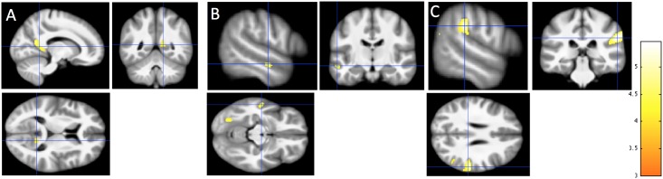
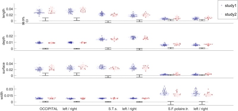
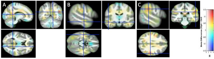
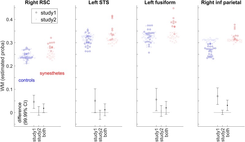
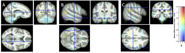
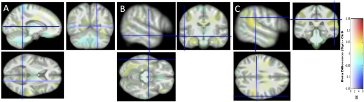
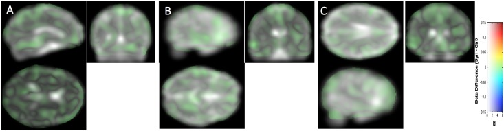
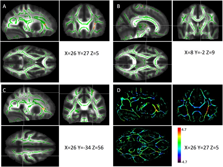
Several publications have reported structural changes in the brain of synesthetes compared to controls, either local differences or differences in connectivity. In the present study, we pursued this quest for structural brain differences that might support the subjective experience of synesthesia. In particular, for the first time in this field, we investigated brain folding in comparing 45 sulcal shapes in each hemisphere of control and grapheme-color synesthete populations. To overcome flaws relative to data interpretation based only on p-values, common in the synesthesia literature, we report confidence intervals of effect sizes. Moreover, our statistical maps are displayed without introducing the classical, but misleading, p-value level threshold. We adopt such a methodological procedure to facilitate appropriate data interpretation and promote the "New Statistics" approach. Based on structural or diffusion magnetic resonance imaging data, we did not find any strong cerebral anomaly, in sulci, tissue volume, tissue density or fiber organization that could support synesthetic color experience. Finally, by sharing our complete datasets, we strongly support the multi-center construction of a sufficient large dataset repository for detecting, if any, subtle brain differences that may help understanding how a subjective experience, such as synesthesia, is mentally constructed.
一些出版物报道了与对照组相比,联觉者大脑的结构变化,无论是局部差异还是连接差异。在本研究中,我们追寻这些可能支持联觉主观体验的结构脑差异。特别是,我们首次在这个领域比较了对照组和字母-颜色联觉者群体的每侧半球的 45 个脑沟形状,以研究脑折叠。为了克服仅基于 p 值的数据分析中存在的缺陷,这种方法在联觉文献中很常见,我们报告了效应大小的置信区间。此外,我们的统计映射是在不引入经典但具有误导性的 p 值水平阈值的情况下显示的。我们采用这种方法来促进适当的数据解释并推广“新统计学”方法。基于结构或扩散磁共振成像数据,我们没有发现任何强烈的大脑异常,无论是在脑沟、组织体积、组织密度还是纤维组织方面,这些都可以支持联觉颜色体验。最后,通过共享我们的完整数据集,我们强烈支持建立一个足够大的多中心数据集存储库,以检测任何可能有助于理解像联觉这样的主观体验是如何在心理上构建的细微大脑差异。