Comprehensive Cancer Center, University of Alabama at Birmingham, Birmingham, AL 35294, USA.
Oncol Rep. 2018 Jun;39(6):2482-2498. doi: 10.3892/or.2018.6332. Epub 2018 Mar 23.
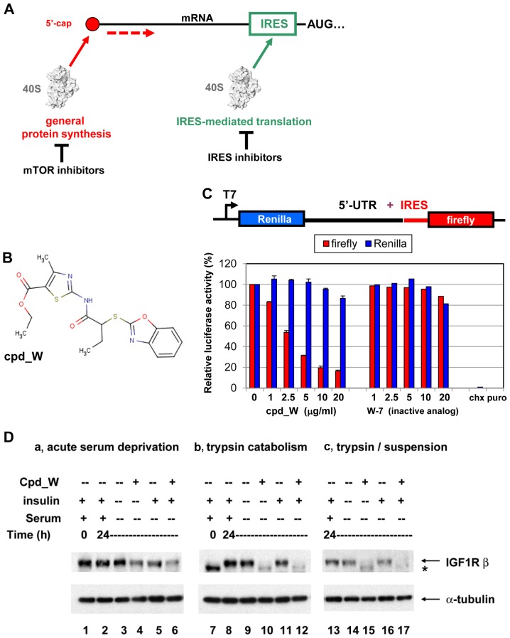
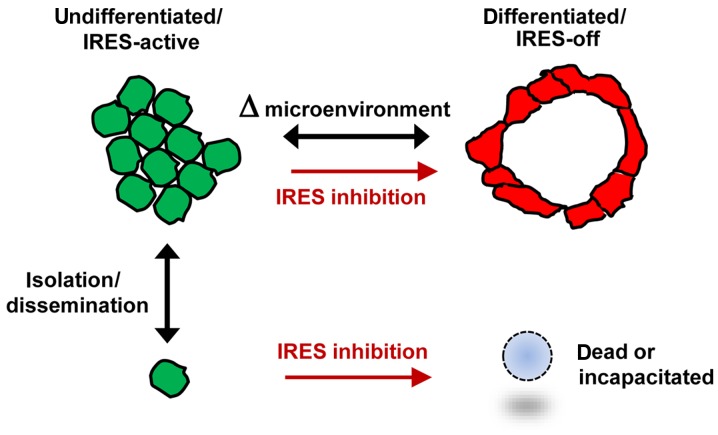
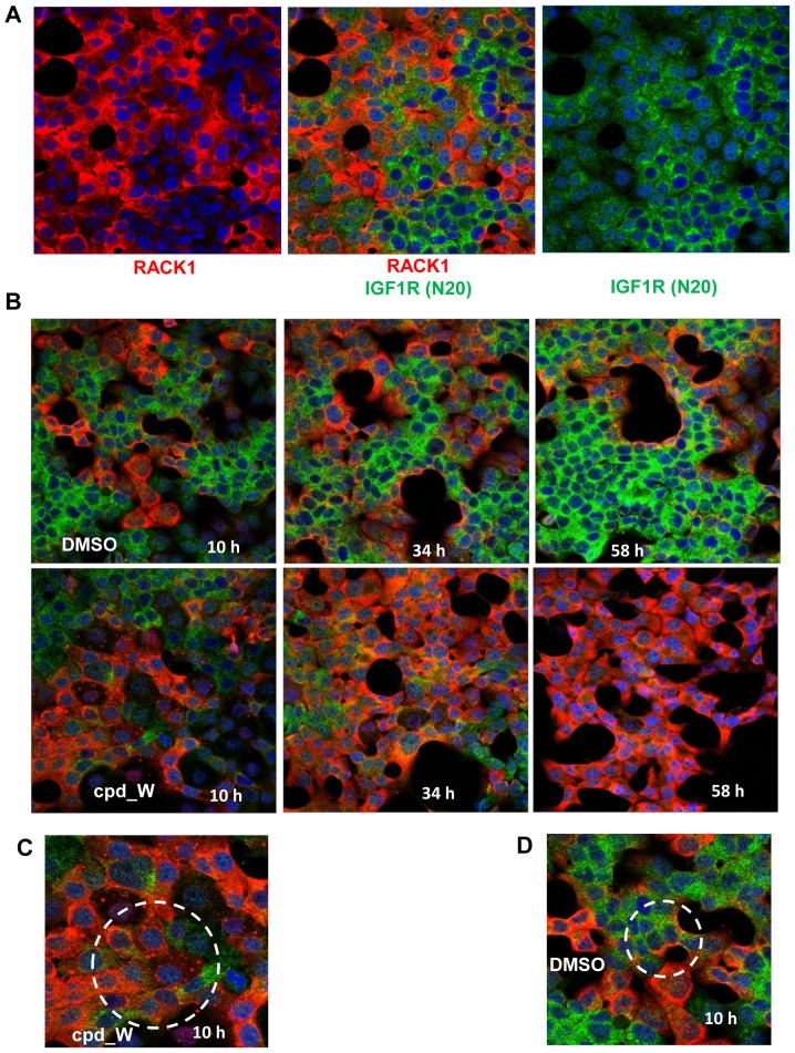
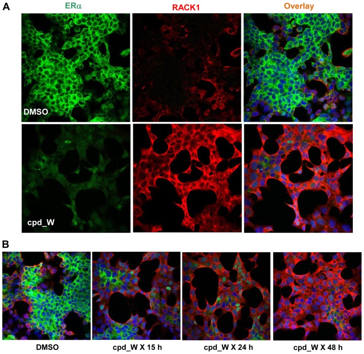
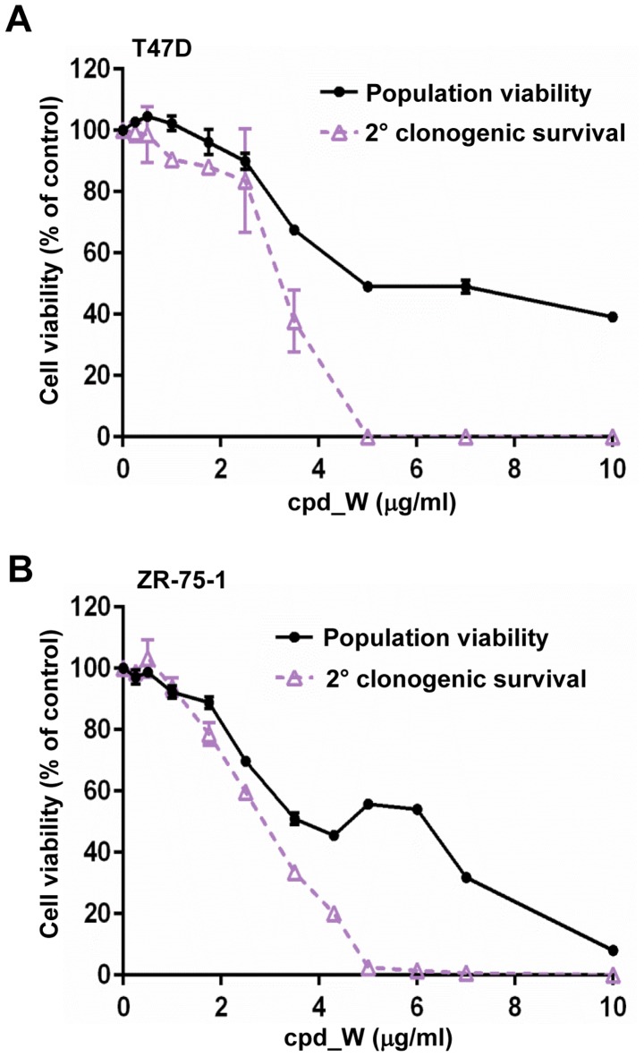
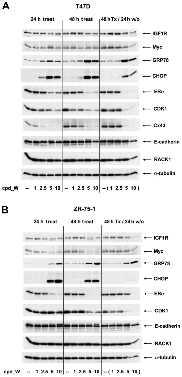
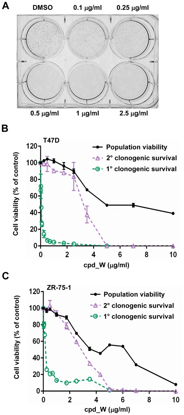
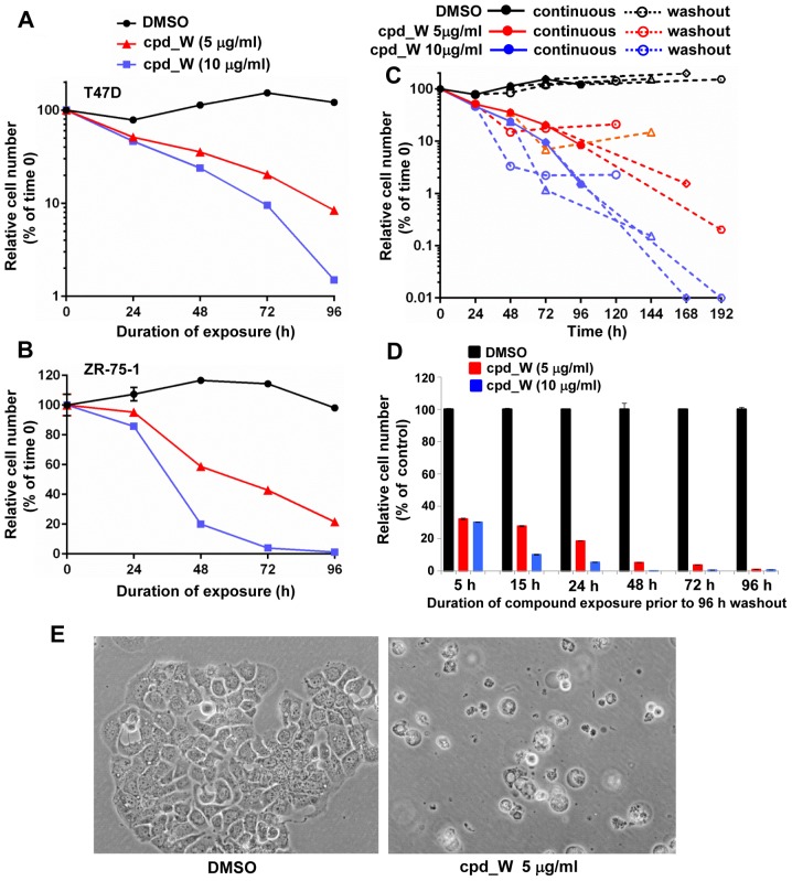
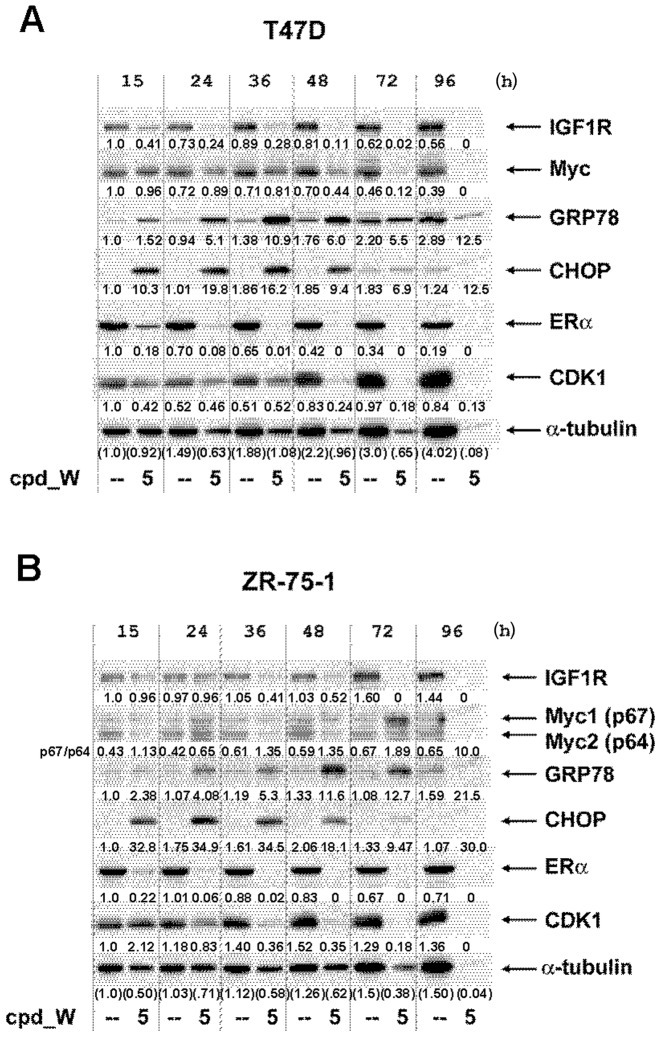
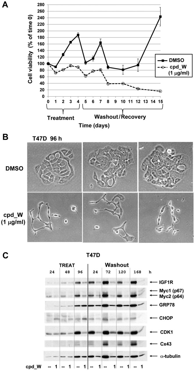
Using a series of potential biomarkers relevant to mechanisms of protein synthesis, we observed that estrogen receptor (ER)-positive breast tumor cells exist in two distinct yet interconvertible phenotypic states (of roughly equal proportion) which differ in the degree of differentiation and use of IRES-mediated translation. Nascently translated IGF1R in the cytoplasm positively correlated with IRES activity and the undifferentiated phenotype, while epitope accessibility of RACK1, an integral component of the 40S ribosomal subunit, aligned with the more differentiated IRES-off state. When deprived of soluble growth factors, the entire tumor cell population shifted to the undifferentiated phenotype in which IRES-mediated translation was active, facilitating survival under these adverse microenvironmental conditions. However, if IRES-mediated translation was inhibited, the cells instead were forced to transition uniformly to the more differentiated state. Notably, cytoplasmic localization of estrogen receptor α (ERα/ESR1) precisely mirrored the pattern observed with nascent IGF1R, correlating with the undifferentiated IRES-active phenotype. Inhibition of IRES-mediated translation resulted in both a shift in ERα to the nucleus (consistent with differentiation) and a marked decrease in ERα abundance (consistent with the inhibition of ERα synthesis via its IRES). Although breast tumor cells tolerated forced differentiation without extensive loss of their viability, their reproductive capacity was severely compromised. In addition, CDK1 was decreased, connexin 43 eliminated and Myc translation altered as a consequence of IRES inhibition. Isolated or low-density ER-positive breast tumor cells were particularly vulnerable to IRES inhibition, losing the ability to generate viable cohesive colonies, or undergoing massive cell death. Collectively, these results provide further evidence for the integral relationship between IRES-mediated translation and the undifferentiated phenotype and demonstrate how therapeutic manipulation of this specialized mode of protein synthesis may be used to limit the phenotypic plasticity and incapacitate or eliminate these otherwise highly resilient breast tumor cells.
利用一系列与蛋白质合成机制相关的潜在生物标志物,我们观察到雌激素受体 (ER)-阳性乳腺癌细胞存在两种截然不同但可相互转化的表型状态(比例大致相等),它们在分化程度和使用 IRES 介导的翻译方面存在差异。细胞质中新生翻译的 IGF1R 与 IRES 活性和未分化表型呈正相关,而 40S 核糖体亚基的组成部分 RACK1 的表位可及性与更分化的 IRES 关闭状态一致。当缺乏可溶性生长因子时,整个肿瘤细胞群转变为未分化表型,其中 IRES 介导的翻译活跃,从而在这些不利的微环境条件下促进生存。然而,如果抑制 IRES 介导的翻译,细胞则被迫统一转变为更分化的状态。值得注意的是,雌激素受体α (ERα/ESR1) 的细胞质定位与新生 IGF1R 观察到的模式精确匹配,与未分化的 IRES 活性表型相关。抑制 IRES 介导的翻译导致 ERα 向核内转移(与分化一致),并且 ERα 丰度显著降低(与通过 IRES 抑制 ERα 合成一致)。尽管乳腺癌细胞耐受强制分化而不会广泛丧失其活力,但它们的繁殖能力受到严重损害。此外,CDK1 减少,连接蛋白 43 消除,Myc 翻译改变是由于 IRES 抑制的结果。分离或低密度 ER 阳性乳腺癌细胞特别容易受到 IRES 抑制的影响,丧失产生有活力的凝聚集落的能力,或发生大量细胞死亡。总的来说,这些结果为 IRES 介导的翻译与未分化表型之间的内在关系提供了进一步的证据,并证明了如何通过治疗性操纵这种特殊的蛋白质合成方式来限制这些表型可塑性并使这些高度有弹性的乳腺癌细胞丧失能力或消除。