Jiangsu Key Laboratory of Oral Diseases, Nanjing Medical University, Nanjing, Jiangsu 210029, P.R. China.
Department of Oral and Maxillofacial Surgery, Ninth People's Hospital, Shanghai 200011, P.R. China.
Oncol Rep. 2018 Jun;39(6):2584-2594. doi: 10.3892/or.2018.6335. Epub 2018 Mar 27.
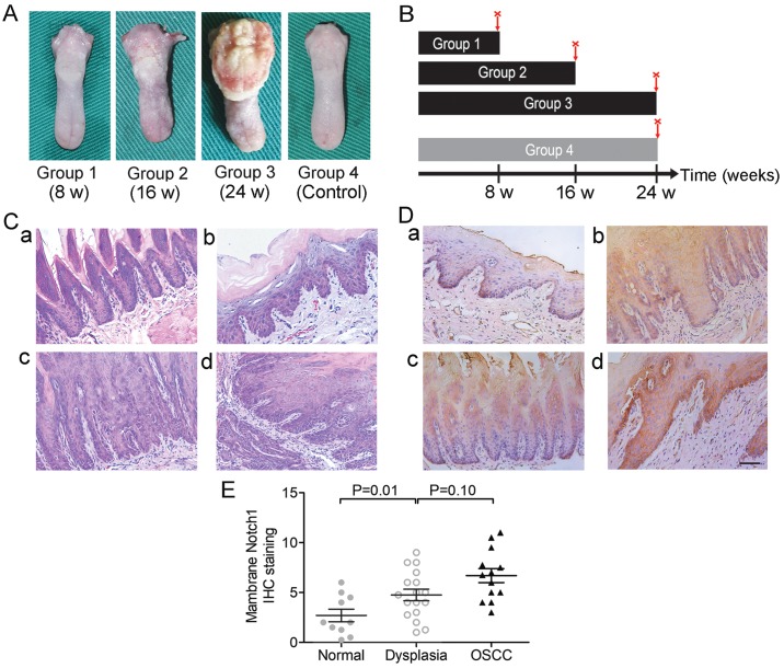
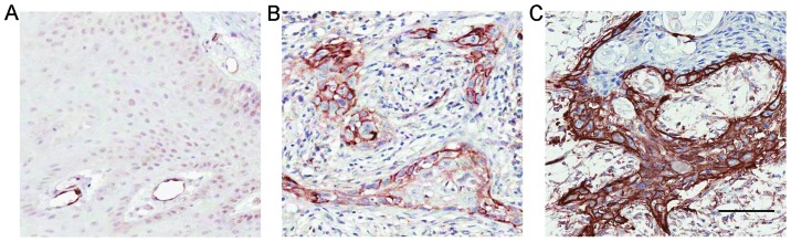
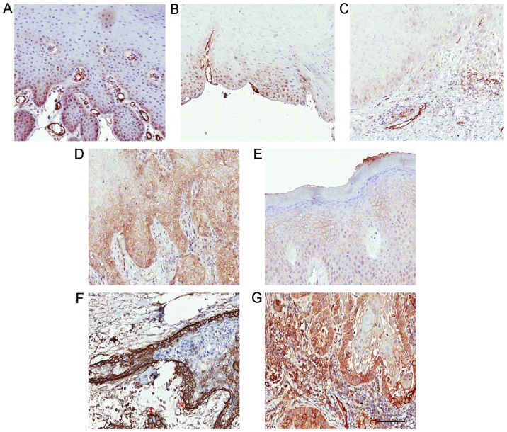
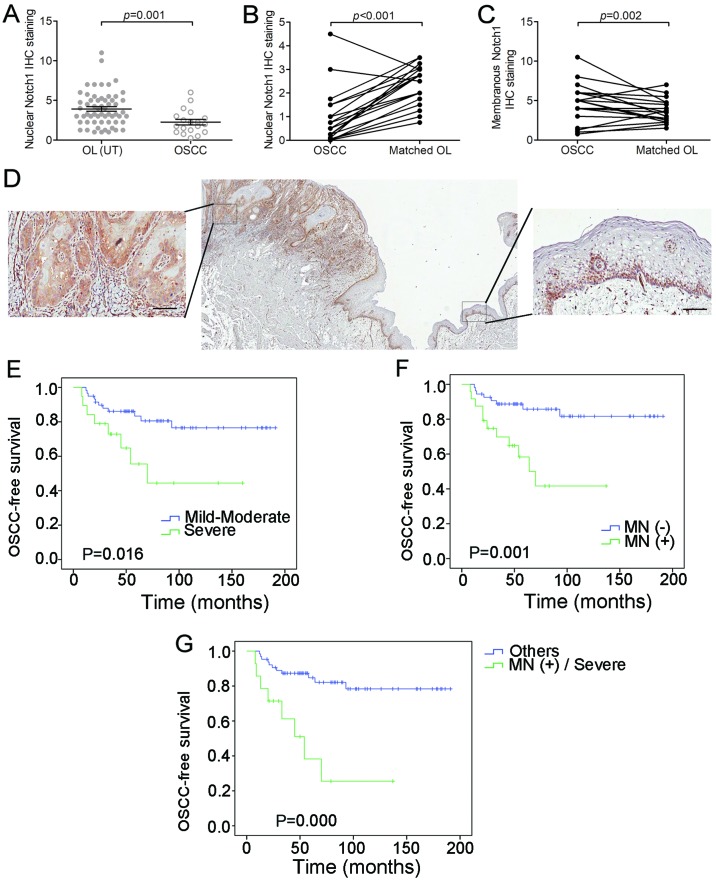
Notch1 signaling is essential for tissue development and tumor progression. This signaling pathway has also been implicated in oral leukoplakia (OL) and oral squamous cell carcinoma (OSCC). However, the role of Notch1 expression in OL and its malignant transformation is unknown. This study aimed to examine the Notch1 expression patterns by immunohistochemistry (IHC) in a cohort of 78 Chinese patients with OL and to analyze the relationship between the patterns and progression of OL to OSCC. Strong Notch1 staining was observed in 10 (13%) of the 78 OL patients, but it was not associated with any of the clinicopathological parameters. However, we observed membranous Notch1 expression in 24 (31%) of the OL samples. Membranous Notch1 expression was significantly associated with the severity of dysplasia (P<0.001) and development of OSCC (P=0.003). By multivariate analysis, membranous Notch1 expression was found to be the only independent factor for OSCC development in the patient population (P=0.019). Among the 24 patients with membranous Notch1 expression, 11 (46%) developed OSCC compared to 8 (15%) of the 54 patients without such expression (P=0.001, determined by log‑rank test). Furthermore, we established a 4‑nitroquinoline‑1‑oxide (4NQO)‑induced murine OSCC model and studied the Notch1 expression patterns in different stages of carcinogenesis. We observed that the extent of expression of membranous Notch1 increased during carcinogenesis. These data indicated a relationship between membranous Notch1 expression and OSCC risk in patients with OL and suggested that membranous Notch1 served as a biomarker for assessing OSCC risk.
Notch1 信号通路对于组织发育和肿瘤进展至关重要。该信号通路也与口腔白斑病 (OL) 和口腔鳞状细胞癌 (OSCC) 有关。然而,Notch1 表达在 OL 及其恶性转化中的作用尚不清楚。本研究旨在通过免疫组织化学 (IHC) 检测 78 例中国 OL 患者的 Notch1 表达模式,并分析其与 OL 向 OSCC 进展的关系。在 78 例 OL 患者中,有 10 例(13%)观察到 Notch1 染色强阳性,但与任何临床病理参数均无相关性。然而,我们在 24 例(31%)OL 样本中观察到膜 Notch1 表达。膜 Notch1 表达与异型增生的严重程度显著相关(P<0.001),与 OSCC 的发生显著相关(P=0.003)。通过多变量分析,膜 Notch1 表达是患者人群中 OSCC 发生的唯一独立因素(P=0.019)。在 24 例膜 Notch1 表达的患者中,有 11 例(46%)发展为 OSCC,而在 54 例无膜 Notch1 表达的患者中,有 8 例(15%)发展为 OSCC(P=0.001,log-rank 检验)。此外,我们建立了 4-硝基喹啉-1-氧化物(4NQO)诱导的小鼠 OSCC 模型,并研究了不同致癌阶段的 Notch1 表达模式。我们观察到膜 Notch1 的表达程度在致癌过程中增加。这些数据表明,OL 患者中膜 Notch1 表达与 OSCC 风险之间存在关联,并提示膜 Notch1 可作为评估 OSCC 风险的生物标志物。