• 文献检索
  • 文档翻译
  • 深度研究
  • 学术资讯
  • Suppr Zotero 插件Zotero 插件
  • 邀请有礼
  • 套餐&价格
  • 历史记录
应用&插件
Suppr Zotero 插件Zotero 插件浏览器插件Mac 客户端Windows 客户端微信小程序
定价
高级版会员购买积分包购买API积分包
服务
文献检索文档翻译深度研究API 文档MCP 服务
关于我们
关于 Suppr公司介绍联系我们用户协议隐私条款
关注我们

Suppr 超能文献

核心技术专利:CN118964589B侵权必究
粤ICP备2023148730 号-1Suppr @ 2026

文献检索

告别复杂PubMed语法,用中文像聊天一样搜索,搜遍4000万医学文献。AI智能推荐,让科研检索更轻松。

立即免费搜索

文件翻译

保留排版,准确专业,支持PDF/Word/PPT等文件格式,支持 12+语言互译。

免费翻译文档

深度研究

AI帮你快速写综述,25分钟生成高质量综述,智能提取关键信息,辅助科研写作。

立即免费体验

氦离子显微镜下细胞和组织中免疫金标记的成像。

Imaging of immunogold labeling in cells and tissues by helium ion microscopy.

机构信息

Biomedical Research Institute, National Institute of Advanced Industrial Science and Technology (AIST), Tsukuba, Ibaraki 305-8566, Japan.

Nanoelectronics Research Institute, National Institute of Advanced Industrial Science and Technology (AIST), Tsukuba, Ibaraki 305-8569, Japan.

出版信息

Int J Mol Med. 2018 Jul;42(1):309-321. doi: 10.3892/ijmm.2018.3604. Epub 2018 Mar 30.

DOI:10.3892/ijmm.2018.3604
PMID:29620251
原文链接:https://pmc.ncbi.nlm.nih.gov/articles/PMC5979831/
Abstract

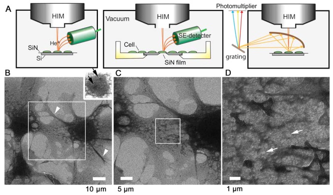
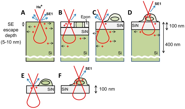
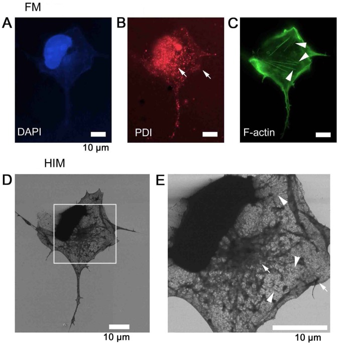
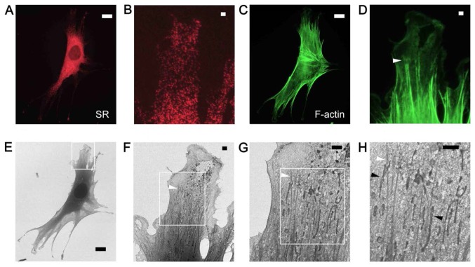
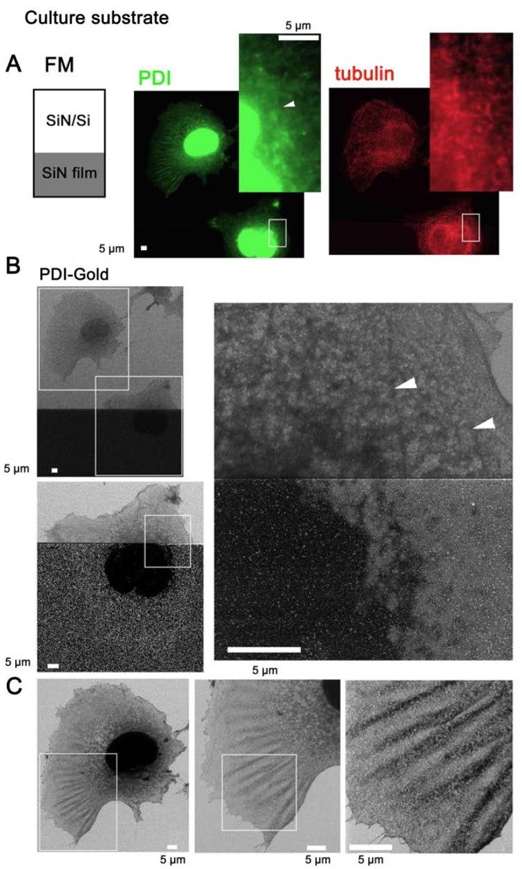
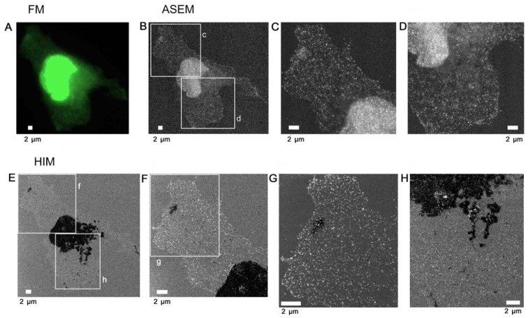
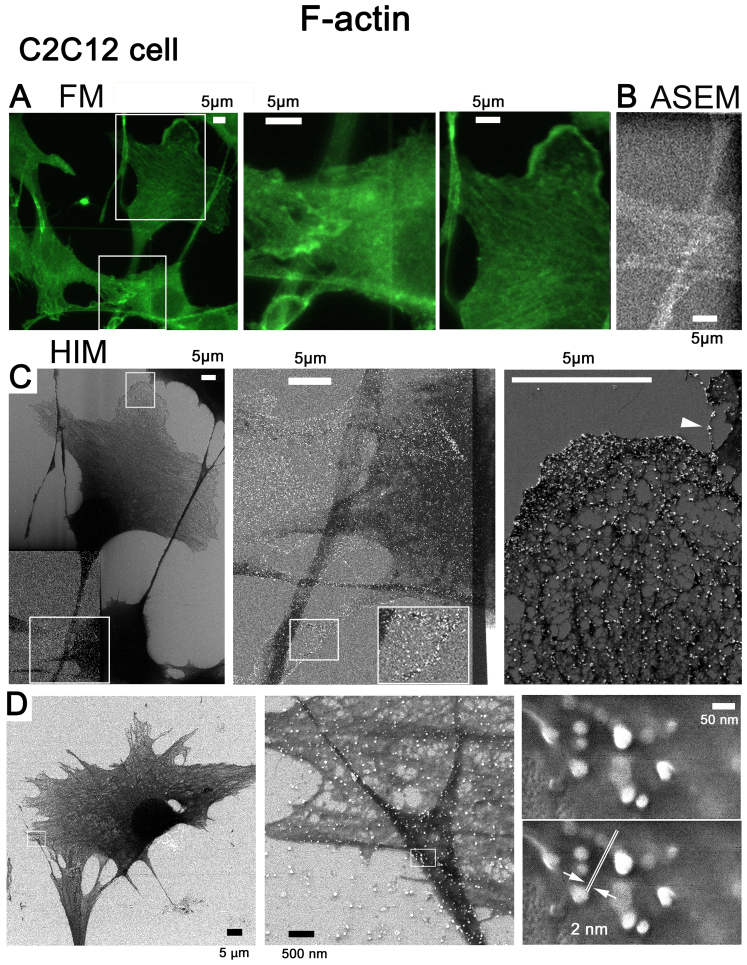
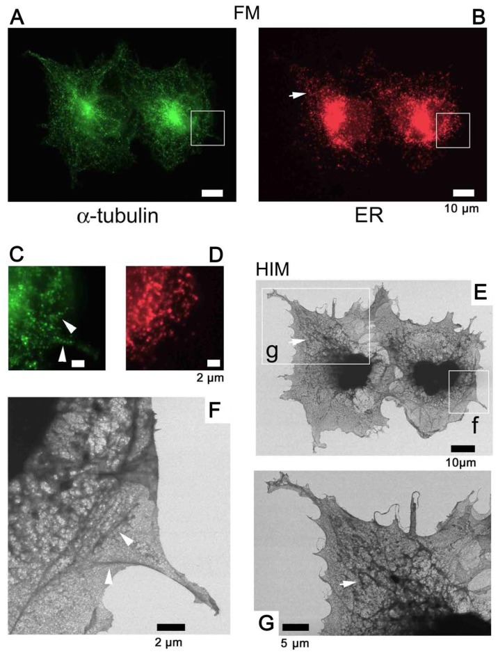
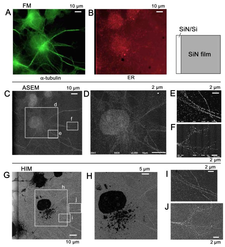
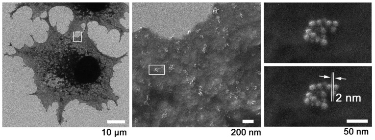
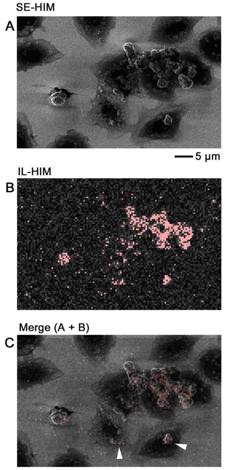
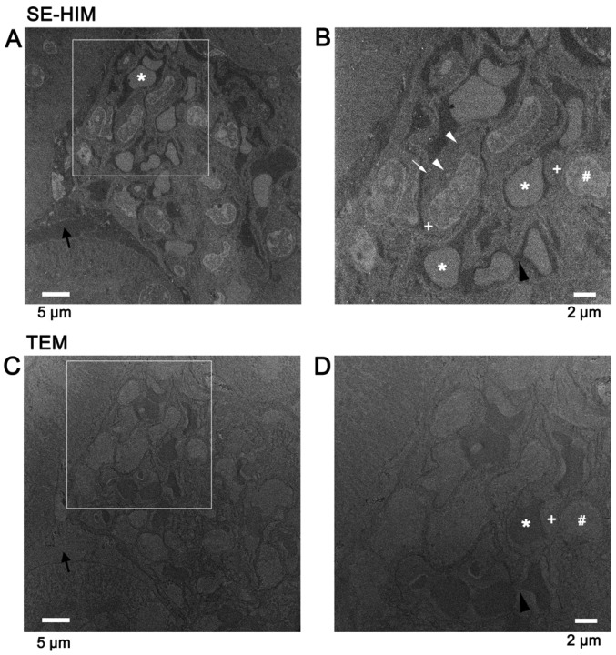
Helium ion microscopy (HIM) scans samples with a fine ion beam exploiting the very short de Broglie wavelength of helium ions. Because the radiation induces only a small sample region to emit secondary electrons (SEs), very high resolution is expected. In order to explore the applications of SE-HIM in biology, COS7 kidney fibroblast cells and C2C12 myoblast cells cultured on a silicon (Si) nitride (SiN)/Si bilayer were dried and directly observed in high vacuum, without coating or staining. High contrast, high depth-of-field images were obtained revealing the nucleus, endoplasmic reticulum, cytoskeleton and putative mitochondria above a bright background from the support. Gold-tagged antibodies were employed to aid organelle identification. Signals from the gold tags were most clearly distinguishable by secondary electron (SE)-HIM when cells were grown on thin SiN film, and the minimum gap measured between gold particles showed the resolution to be 2 nm. Wheat germ agglutinin-gold labeling revealed clusters of gold particles ~50-200 nm in diameter on COS7 cells, which might represent assemblies of glycosylated proteins, suggesting the formation of membrane raft structures that include membrane proteins. SE-HIM also delivered high contrast images of unstained, uncoated, thin sections of Epon‑embedded mouse kidney tissues mounted on a SiN/Si bilayer, revealing the details of sub-tissues and cell organelles. A charge-coupled mechanism explaining the observed SE-HIM contrast is proposed. Ionoluminescence-HIM was also performed targeting zinc oxide particles on cells. In conclusion, the high depth-of-field, high-resolution imaging achieved using HIM may have applications in various fields, including soft materials.

摘要

氦离子显微镜(HIM)利用氦离子的短德布罗意波长精细扫描样品。由于辐射仅使一小部分样品区域发射二次电子(SE),因此预期具有非常高的分辨率。为了探索 SE-HIM 在生物学中的应用,对在氮化硅(SiN)/硅双层上培养的 COS7 肾成纤维细胞和 C2C12 成肌细胞进行干燥,并在高真空条件下直接观察,无需涂层或染色。获得了高对比度、大景深的图像,揭示了细胞核、内质网、细胞骨架和支持物上方明亮背景中的假定线粒体。金标记的抗体被用来帮助识别细胞器。当细胞生长在薄的 SiN 膜上时,金标记物的信号通过二次电子(SE)-HIM 最清晰可辨,测量的金颗粒之间的最小间隙表明分辨率为 2nm。小麦胚凝集素-金标记揭示了 COS7 细胞中直径约 50-200nm 的金颗粒簇,这可能代表糖基化蛋白的聚集,表明包括膜蛋白的膜筏结构的形成。SE-HIM 还对在 SiN/Si 双层上安装的未染色、未涂层的 Epon 包埋的小鼠肾脏组织的薄切片进行了高对比度成像,揭示了亚组织和细胞器官的细节。提出了一种解释观察到的 SE-HIM 对比度的电荷耦合机制。还针对细胞上的氧化锌颗粒进行了离子发光-HIM。总之,使用 HIM 实现的大景深、高分辨率成像可能在包括软材料在内的各个领域有应用。

https://cdn.ncbi.nlm.nih.gov/pmc/blobs/8ef7/5979831/94a18017e094/IJMM-42-01-0309-g12.jpg
https://cdn.ncbi.nlm.nih.gov/pmc/blobs/8ef7/5979831/b165aec809fc/IJMM-42-01-0309-g00.jpg
https://cdn.ncbi.nlm.nih.gov/pmc/blobs/8ef7/5979831/07a6e9b60bcb/IJMM-42-01-0309-g01.jpg
https://cdn.ncbi.nlm.nih.gov/pmc/blobs/8ef7/5979831/6d56030c8dea/IJMM-42-01-0309-g02.jpg
https://cdn.ncbi.nlm.nih.gov/pmc/blobs/8ef7/5979831/98a9eb5f3989/IJMM-42-01-0309-g03.jpg
https://cdn.ncbi.nlm.nih.gov/pmc/blobs/8ef7/5979831/94f0534a7f87/IJMM-42-01-0309-g04.jpg
https://cdn.ncbi.nlm.nih.gov/pmc/blobs/8ef7/5979831/87aff7cf0f40/IJMM-42-01-0309-g05.jpg
https://cdn.ncbi.nlm.nih.gov/pmc/blobs/8ef7/5979831/a14a449def54/IJMM-42-01-0309-g06.jpg
https://cdn.ncbi.nlm.nih.gov/pmc/blobs/8ef7/5979831/2304292e8c40/IJMM-42-01-0309-g07.jpg
https://cdn.ncbi.nlm.nih.gov/pmc/blobs/8ef7/5979831/023941a5f571/IJMM-42-01-0309-g08.jpg
https://cdn.ncbi.nlm.nih.gov/pmc/blobs/8ef7/5979831/872108ccdef3/IJMM-42-01-0309-g09.jpg
https://cdn.ncbi.nlm.nih.gov/pmc/blobs/8ef7/5979831/d5bffdbc3bb0/IJMM-42-01-0309-g10.jpg
https://cdn.ncbi.nlm.nih.gov/pmc/blobs/8ef7/5979831/faf2c1e8f9c9/IJMM-42-01-0309-g11.jpg
https://cdn.ncbi.nlm.nih.gov/pmc/blobs/8ef7/5979831/94a18017e094/IJMM-42-01-0309-g12.jpg

相似文献

1
Imaging of immunogold labeling in cells and tissues by helium ion microscopy.氦离子显微镜下细胞和组织中免疫金标记的成像。
Int J Mol Med. 2018 Jul;42(1):309-321. doi: 10.3892/ijmm.2018.3604. Epub 2018 Mar 30.
2
Reprint of: Atmospheric scanning electron microscope observes cells and tissues in open medium through silicon nitride film.复制品:通过氮化硅膜在开放介质中观察细胞和组织的大气扫描电子显微镜。
J Struct Biol. 2010 Nov;172(2):191-202. doi: 10.1016/j.jsb.2010.08.006. Epub 2010 Aug 17.
3
High resolution helium ion scanning microscopy of the rat kidney.大鼠肾脏的高分辨率氦离子扫描显微镜观察。
PLoS One. 2013;8(3):e57051. doi: 10.1371/journal.pone.0057051. Epub 2013 Mar 7.
4
Ionoluminescence in the helium ion microscope.氦离子显微镜中的离子发光。
Microsc Microanal. 2012 Dec;18(6):1253-62. doi: 10.1017/S1431927612013463. Epub 2012 Dec 14.
5
The effect of magnesium ions on chromosome structure as observed by helium ion microscopy.通过氦离子显微镜观察镁离子对染色体结构的影响。
Microsc Microanal. 2014 Feb;20(1):184-8. doi: 10.1017/S1431927613013792. Epub 2013 Nov 14.
6
Primary cultured neuronal networks and type 2 diabetes model mouse fatty liver tissues in aqueous liquid observed by atmospheric SEM (ASEM): Staining preferences of metal solutions.通过常压扫描电子显微镜(ASEM)观察的原代培养神经元网络和2型糖尿病模型小鼠脂肪肝组织在水性液体中的情况:金属溶液的染色偏好。
Micron. 2019 Mar;118:9-21. doi: 10.1016/j.micron.2018.11.005. Epub 2018 Nov 23.
7
Nanoarchitecture of the ventral disc of Giardia intestinalis as revealed by high-resolution scanning electron microscopy and helium ion microscopy.高分辨率扫描电子显微镜和氦离子显微镜揭示的肠道贾第虫腹吸盘的纳米结构。
Histochem Cell Biol. 2022 Feb;157(2):251-265. doi: 10.1007/s00418-021-02060-2. Epub 2022 Jan 20.
8
Helium Ion Microscopy (HIM) for the imaging of biological samples at sub-nanometer resolution.用于对生物样品进行亚纳米分辨率成像的氦离子显微镜(HIM)。
Sci Rep. 2013 Dec 17;3:3514. doi: 10.1038/srep03514.
9
Immuno EM-OM correlative microscopy in solution by atmospheric scanning electron microscopy (ASEM).溶液中通过大气扫描电子显微镜(ASEM)的免疫电镜-光学相关显微镜。
J Struct Biol. 2012 Nov;180(2):259-70. doi: 10.1016/j.jsb.2012.08.006. Epub 2012 Aug 29.
10
Helium Ion Microscopy Visualizes Lipid Nanodomains in Mammalian Cells.氦离子显微镜可用于观察哺乳动物细胞中的脂质纳米区室。
Small. 2015 Nov 18;11(43):5781-9. doi: 10.1002/smll.201501540. Epub 2015 Oct 5.

引用本文的文献

1
Correlative High-Resolution Imaging of Iron Uptake in Lung Macrophages.肺巨噬细胞中铁摄取的相关高分辨率成像。
Anal Chem. 2022 Sep 20;94(37):12798-12806. doi: 10.1021/acs.analchem.2c02675. Epub 2022 Sep 7.
2
Bio-imaging with the helium-ion microscope: A review.氦离子显微镜的生物成像:综述
Beilstein J Nanotechnol. 2021 Jan 4;12:1-23. doi: 10.3762/bjnano.12.1. eCollection 2021.

本文引用的文献

1
Field-effect detection using phospholipid membranes.使用磷脂膜的场效应检测。
Sci Technol Adv Mater. 2010 Jul 15;11(3):033001. doi: 10.1088/1468-6996/11/3/033001. eCollection 2010 Jun.
2
Observation of tissues in open aqueous solution by atmospheric scanning electron microscopy: applicability to intraoperative cancer diagnosis.通过常压扫描电子显微镜观察开放水溶液中的组织:在术中癌症诊断中的适用性
Int J Oncol. 2015 May;46(5):1872-82. doi: 10.3892/ijo.2015.2905. Epub 2015 Feb 24.
3
High resolution helium ion scanning microscopy of the rat kidney.
大鼠肾脏的高分辨率氦离子扫描显微镜观察。
PLoS One. 2013;8(3):e57051. doi: 10.1371/journal.pone.0057051. Epub 2013 Mar 7.
4
Ionoluminescence in the helium ion microscope.氦离子显微镜中的离子发光。
Microsc Microanal. 2012 Dec;18(6):1253-62. doi: 10.1017/S1431927612013463. Epub 2012 Dec 14.
5
Correlative light and electron microscopy using cathodoluminescence from nanoparticles with distinguishable colours.采用具有可区分颜色的纳米粒子的阴极发光进行相关的光和电子显微镜检查。
Sci Rep. 2012;2:865. doi: 10.1038/srep00865. Epub 2012 Nov 15.
6
Immuno EM-OM correlative microscopy in solution by atmospheric scanning electron microscopy (ASEM).溶液中通过大气扫描电子显微镜(ASEM)的免疫电镜-光学相关显微镜。
J Struct Biol. 2012 Nov;180(2):259-70. doi: 10.1016/j.jsb.2012.08.006. Epub 2012 Aug 29.
7
Whole-cell imaging at nanometer resolutions using fast and slow focused helium ions.利用快速和慢速聚焦氦离子实现纳米分辨率的全细胞成像。
Biophys J. 2011 Oct 5;101(7):1788-93. doi: 10.1016/j.bpj.2011.08.028.
8
Imaging of human colon cancer cells using He-Ion scanning microscopy.应用 He-离子扫描显微镜对人结肠癌细胞成像。
J Microsc. 2011 Jun;242(3):290-4. doi: 10.1111/j.1365-2818.2010.03467.x. Epub 2010 Nov 24.
9
Atmospheric scanning electron microscope observes cells and tissues in open medium through silicon nitride film.大气扫描电子显微镜通过氮化硅膜观察开放介质中的细胞和组织。
J Struct Biol. 2010 Mar;169(3):438-49. doi: 10.1016/j.jsb.2010.01.005. Epub 2010 Jan 15.
10
Contrast mechanisms and image formation in helium ion microscopy.氦离子显微镜中的对比度机制与图像形成
Microsc Microanal. 2009 Apr;15(2):147-53. doi: 10.1017/S1431927609090138.