Novák Dominik, Vadovič Pavol, Ovečka Miroslav, Šamajová Olga, Komis George, Colcombet Jean, Šamaj Jozef
Department of Cell Biology, Centre of the Region Haná for Biotechnological and Agricultural Research, Palacký University Olomouc, Olomouc, Czechia.
UMR9213 Institut des Sciences des Plantes de Paris Saclay, Orsay, France.
Front Plant Sci. 2018 Mar 21;9:371. doi: 10.3389/fpls.2018.00371. eCollection 2018.
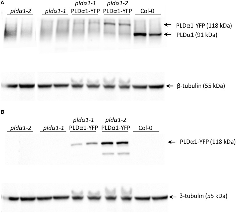
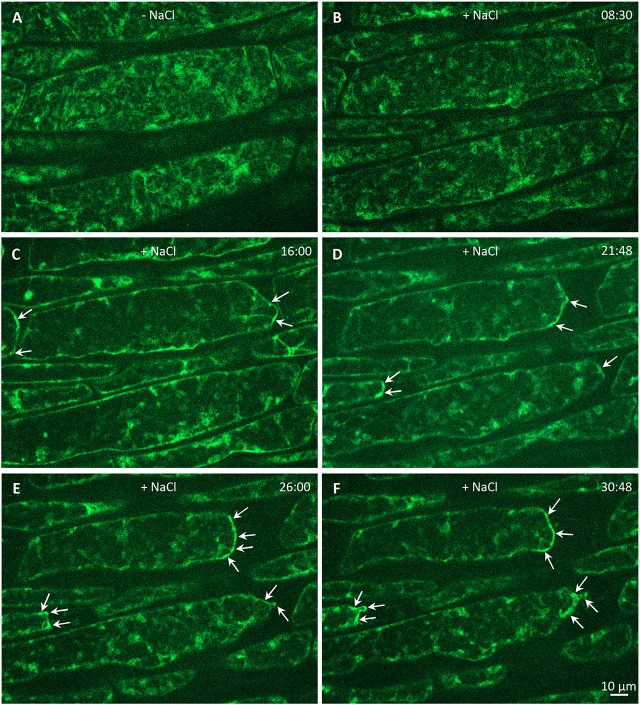
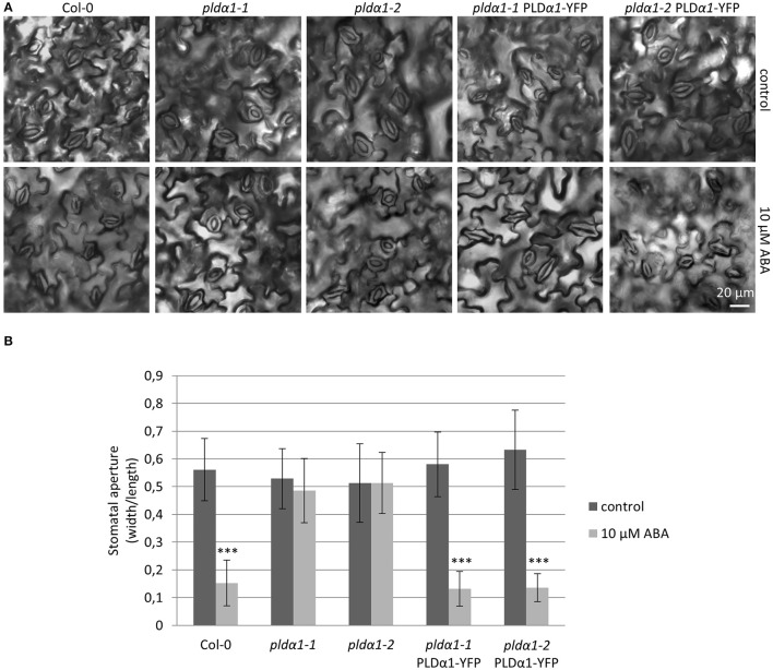
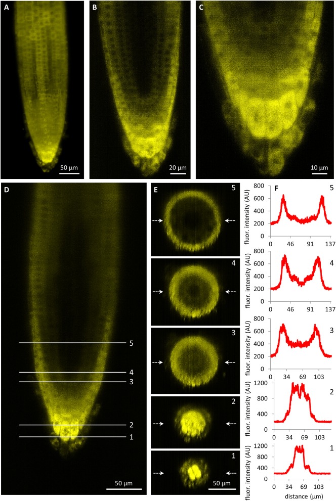
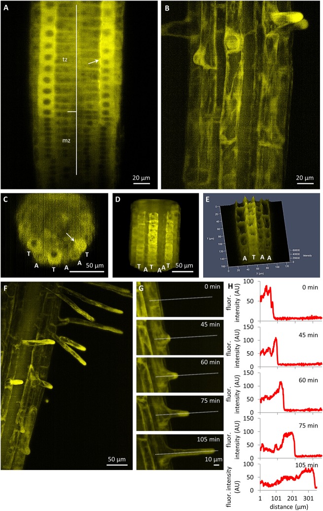
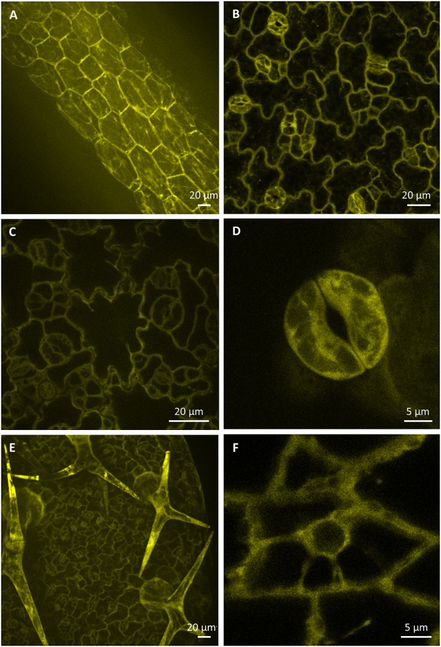
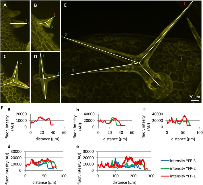
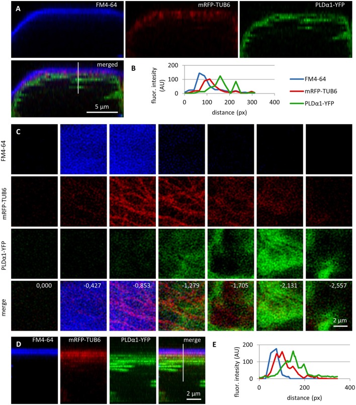
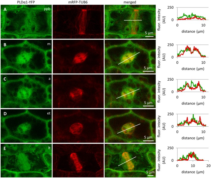
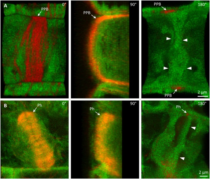
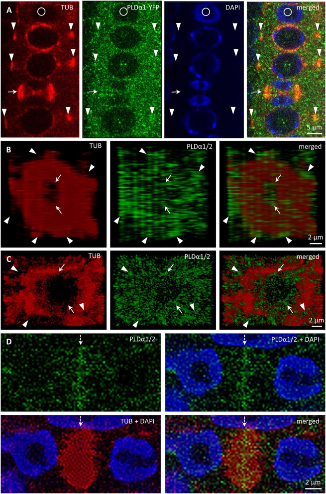
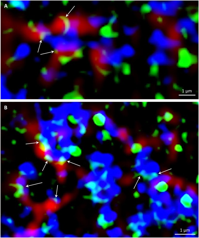
Phospholipase D alpha 1 (PLDα1, At3g15730) and its product phosphatidic acid (PA) are involved in a variety of cellular and physiological processes, such as cytoskeletal remodeling, regulation of stomatal closure and opening, as well as biotic and abiotic stress signaling. Here we aimed to study developmental expression patterns and subcellular localization of PLDα1 in Arabidopsis using advanced microscopy methods such as light-sheet fluorescence microscopy (LSFM) and structured illumination microscopy (SIM). We complemented two knockout mutants with a YFP-tagged PLDα1 expressed under the native promoter in order to study developmental expression pattern and subcellular localization of PLDα1 in under natural conditions. Imaging of tissue-specific and developmentally-regulated localization of YFP-tagged PLDα1 by LSFM in roots of growing seedlings showed accumulation of PLDα1-YFP in the root cap and the rhizodermis. Expression of PLDα1-YFP in the rhizodermis was considerably higher in trichoblasts before and during root hair formation and growth. Thus, PLDα1-YFP accumulated in emerging root hairs and in the tips of growing root hairs. PLDα1-YFP showed cytoplasmic subcellular localization in root cap cells and in cells of the root transition zone. In aerial parts of plants PLDα1-YFP was also localized in the cytoplasm showing enhanced accumulation in the cortical cytoplasmic layer of epidermal non-dividing cells of hypocotyls, leaves, and leaf petioles. However, in dividing cells of root apical meristem and leaf petiole epidermis PLDα1-YFP was enriched in mitotic spindles and phragmoplasts, as revealed by co-visualization with microtubules. Finally, super-resolution SIM imaging revealed association of PLDα1-YFP with both microtubules and clathrin-coated vesicles (CCVs) and pits (CCPs). In conclusion, this study shows the developmentally-controlled expression and subcellular localization of PLDα1 in dividing and non-dividing Arabidopsis cells.
磷脂酶Dα1(PLDα1,At3g15730)及其产物磷脂酸(PA)参与多种细胞和生理过程,如细胞骨架重塑、气孔开闭调节以及生物和非生物胁迫信号传导。在此,我们旨在利用先进的显微镜方法,如光片荧光显微镜(LSFM)和结构光照显微镜(SIM),研究拟南芥中PLDα1的发育表达模式和亚细胞定位。我们用在天然启动子驱动下表达的YFP标记的PLDα1对两个敲除突变体进行互补,以便在自然条件下研究PLDα1的发育表达模式和亚细胞定位。通过LSFM对生长幼苗根部YFP标记的PLDα1进行组织特异性和发育调控定位成像,结果显示PLDα1-YFP在根冠和根表皮中积累。在根毛形成和生长之前及期间,PLDα1-YFP在根表皮毛细胞中的表达明显更高。因此,PLDα1-YFP在新出现的根毛和生长中的根毛尖端积累。PLDα1-YFP在根冠细胞和根过渡区细胞中显示出细胞质亚细胞定位。在植物地上部分,PLDα1-YFP也定位于细胞质中,在下胚轴、叶片和叶柄的表皮非分裂细胞的皮质细胞质层中积累增强。然而,通过与微管共定位发现,在根尖分生组织和叶柄表皮的分裂细胞中,PLDα1-YFP富集于有丝分裂纺锤体和成膜体中。最后,超分辨率SIM成像显示PLDα1-YFP与微管以及网格蛋白包被小泡(CCV)和包被凹陷(CCP)都有关联。总之,本研究展示了PLDα1在拟南芥分裂和非分裂细胞中的发育调控表达及亚细胞定位。