McVitie S, Hughes S, Fallon K, McFadzean S, McGrouther D, Krajnak M, Legrand W, Maccariello D, Collin S, Garcia K, Reyren N, Cros V, Fert A, Zeissler K, Marrows C H
Scottish Universities Physics Alliance, School of Physics and Astronomy, University of Glasgow, Glasgow, G12 8QQ, United Kingdom.
Department of Materials Science and Engineering, Monash University, Clayton, Victoria, 3800, Australia.
Sci Rep. 2018 Apr 9;8(1):5703. doi: 10.1038/s41598-018-23799-0.
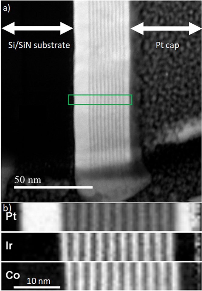
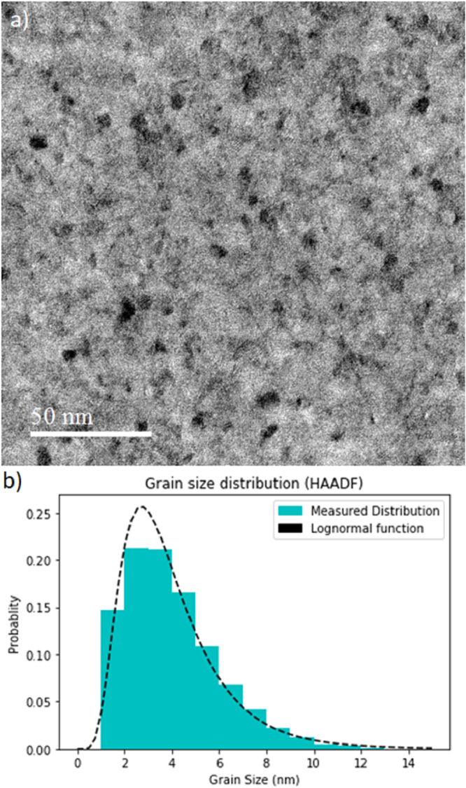
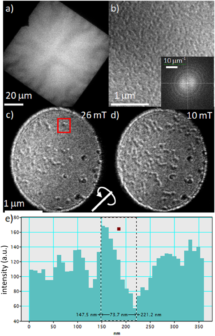
Skyrmions in ultrathin ferromagnetic metal (FM)/heavy metal (HM) multilayer systems produced by conventional sputtering methods have recently generated huge interest due to their applications in the field of spintronics. The sandwich structure with two correctly-chosen heavy metal layers provides an additive interfacial exchange interaction which promotes domain wall or skyrmion spin textures that are Néel in character and with a fixed chirality. Lorentz transmission electron microscopy (TEM) is a high resolution method ideally suited to quantitatively image such chiral magnetic configurations. When allied with physical and chemical TEM analysis of both planar and cross-sectional samples, key length scales such as grain size and the chiral variation of the magnetisation variation have been identified and measured. We present data showing the importance of the grain size (mostly < 10 nm) measured from direct imaging and its potential role in describing observed behaviour of isolated skyrmions (diameter < 100 nm). In the latter the region in which the magnetization rotates is measured to be around 30 nm. Such quantitative information on the multiscale magnetisation variations in the system is key to understanding and exploiting the behaviour of skyrmions for future applications in information storage and logic devices.
通过传统溅射方法制备的超薄铁磁金属(FM)/重金属(HM)多层系统中的斯格明子,因其在自旋电子学领域的应用,最近引起了极大的关注。具有两个正确选择的重金属层的三明治结构提供了一种附加的界面交换相互作用,这种相互作用促进了具有奈尔特性和固定手性的畴壁或斯格明子自旋纹理。洛伦兹透射电子显微镜(TEM)是一种高分辨率方法,非常适合对这种手性磁结构进行定量成像。当与平面和横截面样品的物理和化学TEM分析相结合时,已经确定并测量了诸如晶粒尺寸和磁化变化的手性变化等关键长度尺度。我们展示的数据表明了从直接成像测量的晶粒尺寸(大多<10nm)的重要性及其在描述孤立斯格明子(直径<100nm)的观测行为中的潜在作用。在后者中,磁化旋转的区域测量为约30nm。系统中多尺度磁化变化的此类定量信息是理解和利用斯格明子行为以用于未来信息存储和逻辑器件应用的关键。