Department of Radiation OncologyUniversity of Kansas Medical Center, Kansas City, Kansas, USA.
Department of Anatomy and Cell BiologyUniversity of Kansas Medical Center, Kansas City, Kansas, USA.
Reproduction. 2018 Jun;155(6):553-562. doi: 10.1530/REP-18-0089. Epub 2018 Apr 10.
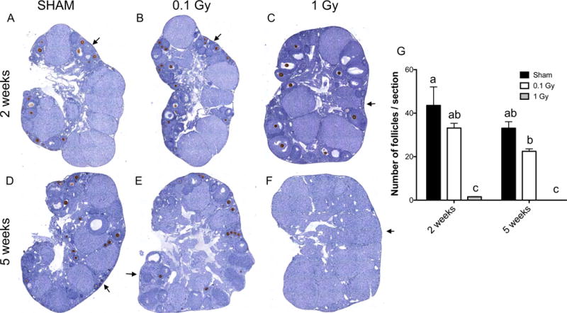
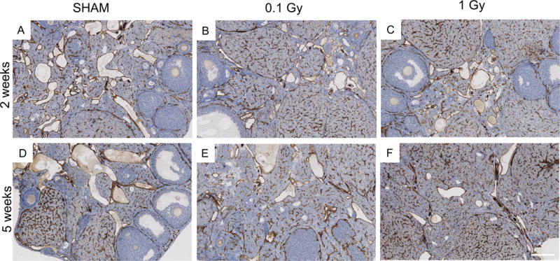
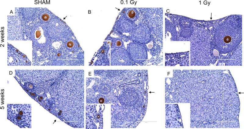
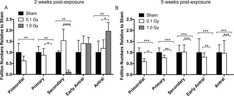
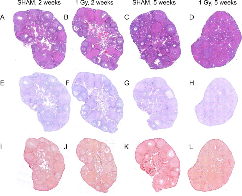
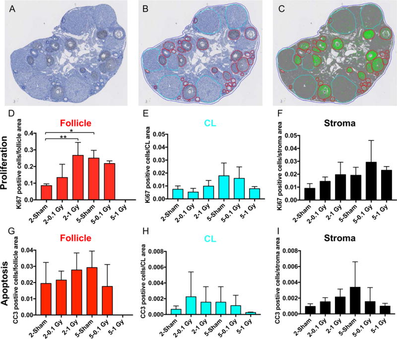
Radiation damage due to total body irradiation (TBI) or targeted abdominal radiation can deplete ovarian follicles and accelerate reproductive aging. We characterized a mouse model of low-dose TBI to investigate how radiation affects the follicular and stromal compartments of the ovary. A single TBI dose of either 0.1 Gy or 1 Gy (Cesium-137 γ) was delivered to reproductively adult CD1 female mice, and sham-treated mice served as controls. Mice were euthanized either 2 weeks or 5 weeks post exposure, and ovarian tissue was harvested. To assess the ovarian reserve, we classified and counted the number of morphologically normal follicles in ovarian histologic sections for all experimental cohorts using an objective method based on immunohistochemistry for an oocyte-specific protein (MSY2). 0.1 Gy did not affect that total number of ovarian follicles, whereas 1 Gy resulted in a dramatic loss. At two weeks, there was a significant reduction in all preantral follicles, but early antral and antral follicles were still present. By five weeks, there was complete depletion of all follicle classes. We examined stromal quality using histologic stains to visualize ovarian architecture and fibrosis and by immunohistochemistry and quantitative microscopy to assess cell proliferation, cell death and vasculature. There were no differences in the ovarian stroma across cohorts with respect to these markers, indicating that this compartment is more radio-resistant relative to the germ cells. These findings have implications for reproductive health and the field of fertility preservation because the radiation doses we examined mimic scatter doses experienced in typical therapeutic regimens.
全身照射(TBI)或靶向腹部放射治疗引起的辐射损伤会耗尽卵巢卵泡并加速生殖衰老。我们描述了一种低剂量 TBI 的小鼠模型,以研究辐射如何影响卵巢的卵泡和基质区室。生殖成熟的 CD1 雌性小鼠接受单次 TBI 剂量 0.1Gy 或 1Gy(铯-137γ)照射,假照射小鼠作为对照。照射后 2 周或 5 周处死小鼠,采集卵巢组织。为了评估卵巢储备,我们使用基于卵母细胞特异性蛋白(MSY2)免疫组化的客观方法对所有实验组的卵巢组织切片进行分类并计数形态正常的卵泡数量。0.1Gy 不影响卵巢卵泡总数,而 1Gy 则导致明显损失。2 周时,所有原始卵泡数量显著减少,但仍存在早期窦前卵泡和窦状卵泡。5 周时,所有卵泡均完全耗尽。我们使用组织学染色观察卵巢结构和纤维化,以及免疫组织化学和定量显微镜检查来评估细胞增殖、细胞死亡和血管生成,以此来检查基质质量。在这些标志物方面,各队列之间的卵巢基质没有差异,这表明与生殖细胞相比,该区室具有更高的辐射抗性。这些发现对生殖健康和生育力保存领域具有重要意义,因为我们研究的辐射剂量模拟了典型治疗方案中所经历的散射剂量。