1 Banque de Tissus et de Cellules des Hospices Civils de Lyon, Hôpital Edouard Herriot, Lyon, France.
2 Université Claude Bernard Lyon I, Villeurbanne, Lyon, France.
Cell Transplant. 2018 Feb;27(2):264-274. doi: 10.1177/0963689717741140.
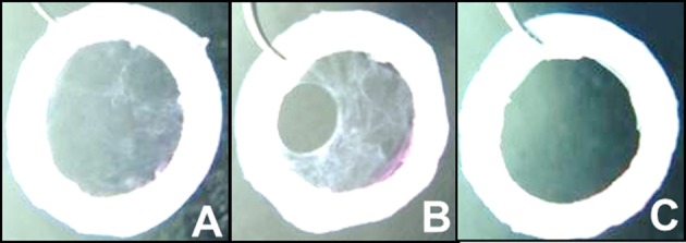
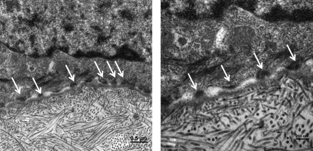
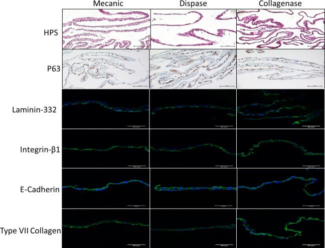
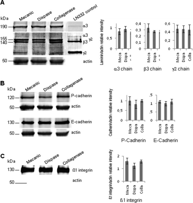
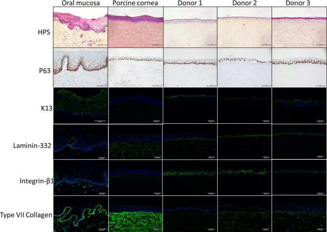
Total bilateral limbal stem cell deficiency leading to loss of corneal clarity, potential vision loss, pain, photophobia, and keratoplasty failure cannot be treated by autologous limbal transplantation, and allogeneic limbal transplantation requires subsequent immunosuppressive treatment. Cultured autologous oral mucosal epithelial cells have been shown to be safe and effective alternatives. These cells can be transplanted on supports or without support after detachment from the culture dishes. Dispase, known for epidermal sheet detachment, is reported as not usable for oral mucosa. The objective was to find an optimized detachment method providing a sufficiently resistant and adhesive cultured oral mucosal epithelium (COME), which can be grafted without sutures. Enzymatic treatments (dispase or collagenase at different concentrations) were compared to enzyme-free mechanical detachment. Histological immunofluorescence (IF) and Western blotting (WB) were used to examine the impact on adhesion markers (laminin-332, β1-integrin, and type VII collagen) and junctional markers (E-cadherin, P-cadherin). Finally, the COME ability to adhere to the cornea and produce a differentiated epithelium 15 d after grafting onto an ex vivo porcine stroma model were investigated by histology, IF, and transmission electron microscopy. Collagenase at 0.5 mg/mL and dispase at 5 mg/mL were selected for comparative study on adhesive expression marker by IF and WB showed that levels of basement membrane proteins and cell-cell and cell-matrix junction proteins were not significantly different between the 3 detachment methods. Collagenase 0.5 mg/mL was selected for the next step validation because of the better reproducibility, 100% success (vs. 33% with dispase 5 mg/mL). Grafted onto porcine de-epithelialized corneal stroma, collagenase 0.5 mg/mL detached COME were found to adhere, stratify, and continue to ensure renewal of the epithelium. For COME, collagenase 0.5 mg/mL enzymatic detachment was selected and validated on its resistance and adhesive marker expression as well as their anchorage onto our new ex vivo de-epithelialized stroma model.
双侧角膜缘干细胞缺乏导致角膜混浊、潜在视力丧失、疼痛、畏光和角膜移植失败,不能通过自体角膜缘移植治疗,同种异体角膜缘移植需要后续免疫抑制治疗。已证明培养的自体口腔黏膜上皮细胞是安全有效的替代物。这些细胞可以在从培养皿中分离后在载体上或无载体的情况下进行移植。已知用于表皮片分离的Dispase 不适用于口腔黏膜。目的是找到一种优化的分离方法,提供足够坚固和黏附的培养口腔黏膜上皮细胞(COME),无需缝合即可移植。比较了酶处理(不同浓度的 Dispase 或胶原酶)和无酶机械分离。组织学免疫荧光(IF)和 Western blot(WB)用于检查对黏附标志物(层粘连蛋白-332、β1 整联蛋白和 VII 型胶原)和连接标志物(E-钙黏蛋白、P-钙黏蛋白)的影响。最后,通过组织学、IF 和透射电子显微镜检查 COME 在移植到体外猪基质模型 15 天后黏附角膜并产生分化上皮的能力。选择胶原酶 0.5mg/mL 和 Dispase 5mg/mL 用于 IF 和 WB 比较黏附表达标志物的研究,结果显示 3 种分离方法之间基底膜蛋白和细胞-细胞和细胞-基质连接蛋白的水平没有显著差异。由于更好的可重复性,选择胶原酶 0.5mg/mL 用于下一步验证,成功率为 100%(Dispase 5mg/mL 为 33%)。将胶原酶 0.5mg/mL 分离的 COME 移植到猪去上皮角膜基质上,发现其能黏附、分层并继续确保上皮更新。对于 COME,选择胶原酶 0.5mg/mL 酶分离,并对其抵抗性和黏附标志物表达及其在我们新的体外去上皮化基质模型上的锚定进行验证。