• 文献检索
  • 文档翻译
  • 深度研究
  • 学术资讯
  • Suppr Zotero 插件Zotero 插件
  • 邀请有礼
  • 套餐&价格
  • 历史记录
应用&插件
Suppr Zotero 插件Zotero 插件浏览器插件Mac 客户端Windows 客户端微信小程序
定价
高级版会员购买积分包购买API积分包
服务
文献检索文档翻译深度研究API 文档MCP 服务
关于我们
关于 Suppr公司介绍联系我们用户协议隐私条款
关注我们

Suppr 超能文献

核心技术专利:CN118964589B侵权必究
粤ICP备2023148730 号-1Suppr @ 2026

文献检索

告别复杂PubMed语法,用中文像聊天一样搜索,搜遍4000万医学文献。AI智能推荐,让科研检索更轻松。

立即免费搜索

文件翻译

保留排版,准确专业,支持PDF/Word/PPT等文件格式,支持 12+语言互译。

免费翻译文档

深度研究

AI帮你快速写综述,25分钟生成高质量综述,智能提取关键信息,辅助科研写作。

立即免费体验

经相位对比磁共振成像术对颈动脉支架置入术前和术后的多次血流测量:一项观察性研究。

Multiple blood flow measurements before and after carotid artery stenting via phase-contrast magnetic resonance imaging: An observational study.

机构信息

Department of Radiology, Osaka University Graduate School of Medicine, Suita, Osaka, Japan.

Department of Neurosurgery, Osaka University Graduate School of Medicine, Suita, Osaka, Japan.

出版信息

PLoS One. 2018 Apr 11;13(4):e0195099. doi: 10.1371/journal.pone.0195099. eCollection 2018.

DOI:10.1371/journal.pone.0195099
PMID:29641548
原文链接:https://pmc.ncbi.nlm.nih.gov/articles/PMC5895018/
Abstract

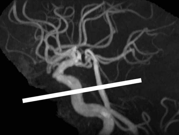
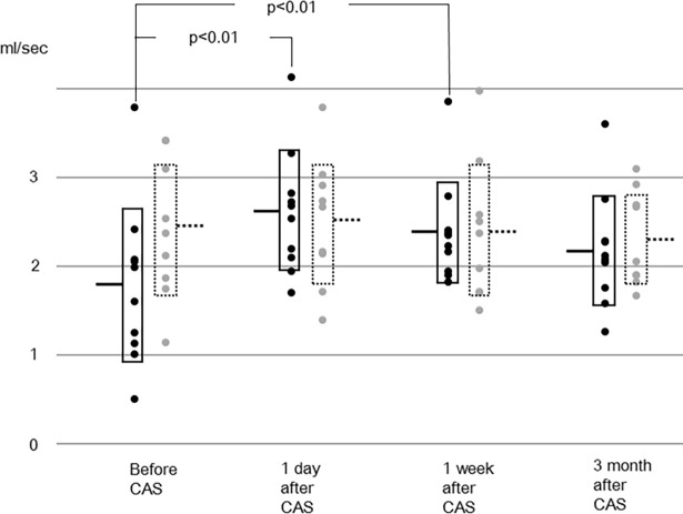
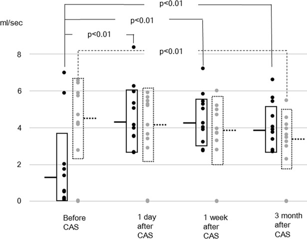
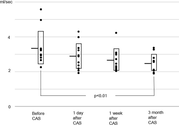
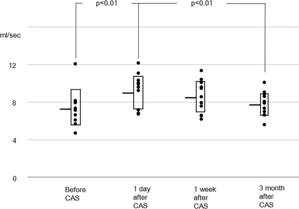
After carotid artery stenting, the procurement of information about blood flow redistribution among brain-feeding arteries and its time trend is essential to understanding a patient's physiological background and to determine their care regimen. Cerebral blood flow has been measured twice following carotid artery stenting in few previous studies, with some discrepancies in the results. The purpose of this study was to measure cerebral blood flow at multiple time points after carotid artery stenting, and to elucidate the time trend of cerebral blood flow and redistribution among arteries. Blood flow rates in 11 subjects were measured preoperatively, at one day, one week, and about three months, respectively after carotid artery stenting by using phase-contrast magnetic resonance imaging. The target vessels were the bilateral internal carotid arteries, the basilar artery, and the bilateral middle cerebral arteries. Lumen was semi-automatically defined using an algorithm utilizing pulsatility. The results showed that blood flow rates in the stented internal carotid artery and the ipsilateral middle cerebral artery increased following carotid artery stenting. Blood flow rates in the contralateral internal carotid artery and the basilar artery gradually declined, and they were lower than the preoperative values at three months after stenting. The sum of blood flow rates of the bilateral internal carotid arteries and the basilar artery increased after carotid artery stenting, and then decreased over the next three months. There was no significant change in the blood flow rate in the contralateral middle cerebral artery. From these results, it was concluded that redistribution among the bilateral internal carotid arteries and the basilar artery occurs after carotid artery stenting, and that it takes months thereafter to reach another equilibrium.

摘要

在颈动脉支架置入术后,了解脑供血动脉之间的血流再分布及其时间趋势对于理解患者的生理背景和确定其护理方案至关重要。在之前的一些研究中,仅对颈动脉支架置入术后的少数患者进行了两次脑血流测量,结果存在一些差异。本研究的目的是在颈动脉支架置入术后的多个时间点测量脑血流,并阐明脑血流和动脉再分布的时间趋势。通过相位对比磁共振成像,在术前、术后 1 天、1 周和大约 3 个月时分别测量了 11 名受试者的血流速度。目标血管为双侧颈内动脉、基底动脉和双侧大脑中动脉。使用利用脉动性的算法半自动定义管腔。结果表明,颈动脉支架置入后,支架内颈内动脉和同侧大脑中动脉的血流速度增加。对侧颈内动脉和基底动脉的血流速度逐渐下降,且在支架置入 3 个月后低于术前值。双侧颈内动脉和基底动脉的血流速度总和在颈动脉支架置入后增加,随后在接下来的 3 个月内下降。对侧大脑中动脉的血流速度没有明显变化。从这些结果可以得出结论,颈动脉支架置入后双侧颈内动脉和基底动脉之间发生了再分布,此后需要数月才能达到另一个平衡。

https://cdn.ncbi.nlm.nih.gov/pmc/blobs/a100/5895018/0bea3a44bdb0/pone.0195099.g005.jpg
https://cdn.ncbi.nlm.nih.gov/pmc/blobs/a100/5895018/863e14a18bd9/pone.0195099.g001.jpg
https://cdn.ncbi.nlm.nih.gov/pmc/blobs/a100/5895018/74d38ce0b9c1/pone.0195099.g002.jpg
https://cdn.ncbi.nlm.nih.gov/pmc/blobs/a100/5895018/56d9708d7e9d/pone.0195099.g003.jpg
https://cdn.ncbi.nlm.nih.gov/pmc/blobs/a100/5895018/62f9b40f5e7d/pone.0195099.g004.jpg
https://cdn.ncbi.nlm.nih.gov/pmc/blobs/a100/5895018/0bea3a44bdb0/pone.0195099.g005.jpg

相似文献

1
Multiple blood flow measurements before and after carotid artery stenting via phase-contrast magnetic resonance imaging: An observational study.经相位对比磁共振成像术对颈动脉支架置入术前和术后的多次血流测量:一项观察性研究。
PLoS One. 2018 Apr 11;13(4):e0195099. doi: 10.1371/journal.pone.0195099. eCollection 2018.
2
Flow volume changes in the major cerebral arteries before and after carotid endarterectomy: an MR angiography study.颈动脉内膜切除术前、后大脑主要动脉的血流容积变化:一项磁共振血管造影研究。
Eur J Vasc Endovasc Surg. 1997 Dec;14(6):446-50. doi: 10.1016/s1078-5884(97)80122-0.
3
Relationship between variations in the circle of Willis and flow rates in internal carotid and basilar arteries determined by means of magnetic resonance imaging with semiautomated lumen segmentation: reference data from 125 healthy volunteers.通过半自动管腔分割的磁共振成像确定 Willis 环变异与颈内动脉和基底动脉血流速度之间的关系:来自 125 名健康志愿者的参考数据
AJNR Am J Neuroradiol. 2006 Sep;27(8):1770-5.
4
TCD evaluation before and after stenting in patients with severe primary carotid artery stenosis versus restenosis.严重原发性颈动脉狭窄与再狭窄患者支架置入前后的经颅多普勒超声(TCD)评估
J Endovasc Ther. 2007 Aug;14(4):483-8. doi: 10.1177/152660280701400408.
5
Effect of internal carotid artery stenting on superior thyroid artery Doppler flow.颈内动脉支架置入术对甲状腺上动脉多普勒血流的影响。
J Ultrasound Med. 2014 Oct;33(10):1783-9. doi: 10.7863/ultra.33.10.1783.
6
Cerebral blood flow and oxygen metabolism measurements using positron emission tomography on the first day after carotid artery stenting.经颅多普勒超声对颈动脉支架置入术后第一天脑血流和氧代谢的测量。
J Stroke Cerebrovasc Dis. 2014 Feb;23(2):e55-64. doi: 10.1016/j.jstrokecerebrovasdis.2013.08.012. Epub 2013 Oct 9.
7
Internal carotid artery stenosis with persistent primitive hypoglossal artery treated with carotid artery stenting: A case report and literature review.经颈动脉支架置入术治疗的伴有永存舌下动脉的颈内动脉狭窄:一例病例报告及文献综述
Neuroradiol J. 2016 Apr;29(2):115-21. doi: 10.1177/1971400915626427. Epub 2016 Jan 29.
8
Blood flow volume quantification of cerebral ischemia: comparison of three noninvasive imaging techniques of carotid and vertebral arteries.脑缺血血流容积定量:三种颈动脉和椎动脉无创成像技术的比较
AJR Am J Roentgenol. 2002 Mar;178(3):551-6. doi: 10.2214/ajr.178.3.1780551.
9
The paradoxical flow hypothesis of the carotid artery: supporting evidence from phase-contrast magnetic resonance imaging.颈动脉矛盾血流假说:来自相位对比磁共振成像的支持证据。
J Stroke Cerebrovasc Dis. 2008 Mar-Apr;17(2):101-8. doi: 10.1016/j.jstrokecerebrovasdis.2007.10.004.
10
Computed tomography indicators of cerebral microperfusion improve long term after carotid stenting in symptomatic patients.有症状患者行颈动脉支架置入术后,脑微灌注的计算机断层扫描指标长期改善。
Acta Biochim Pol. 2019 Apr 8;66(2):229-236. doi: 10.18388/abp.2018_2793.

引用本文的文献

1
Age-Related Tortuosity of Carotid and Vertebral Arteries: Quantitative Evaluation With MR Angiography.年龄相关的颈动脉和椎动脉迂曲:磁共振血管造影定量评估
Front Neurol. 2022 Apr 29;13:858805. doi: 10.3389/fneur.2022.858805. eCollection 2022.
2
Common Carotid Artery Volume Flow: A Comparison Study between Ultrasound Vector Flow Imaging and Phase Contrast Magnetic Resonance Imaging.颈总动脉容积血流:超声矢量血流成像与相位对比磁共振成像的比较研究
Neurol Int. 2021 Jun 23;13(3):269-278. doi: 10.3390/neurolint13030028.
3
Anatomical Considerations for Endovascular Intervention for Extracranial Carotid Disease: A Review of the Literature and Recommended Guidelines.

本文引用的文献

1
How asymptomatic is "asymptomatic" carotid stenosis? Resolving fundamental confusion(s) - and confusions yet to be resolved.“无症状的”颈动脉狭窄究竟有多无症状?解决基本的困惑——以及有待解决的困惑。
Pol Arch Intern Med. 2017 Nov 30;127(11):718-719. doi: 10.20452/pamw.4157.
2
Effect of carotid endarterectomy on retinal function in asymptomatic patients with hemodynamically significant carotid artery stenosis.颈动脉内膜切除术对有症状性颈动脉狭窄患者视网膜功能的影响。
Pol Arch Intern Med. 2017 Nov 30;127(11):722-729. doi: 10.20452/pamw.4064. Epub 2017 Jul 19.
3
Indocyanine green kinetics with near-infrared spectroscopy predicts cerebral hyperperfusion syndrome after carotid artery stenting.
颅外颈动脉疾病血管内介入治疗的解剖学考量:文献综述与推荐指南
J Clin Med. 2020 Oct 27;9(11):3460. doi: 10.3390/jcm9113460.
吲哚菁绿动力学结合近红外光谱可预测颈动脉支架置入术后的脑过度灌注综合征。
PLoS One. 2017 Jul 12;12(7):e0180684. doi: 10.1371/journal.pone.0180684. eCollection 2017.
4
Carotid Artery Stenting and Blood-Brain Barrier Permeability in Subjects with Chronic Carotid Artery Stenosis.慢性颈动脉狭窄患者的颈动脉支架置入术与血脑屏障通透性
Int J Mol Sci. 2017 May 8;18(5):1008. doi: 10.3390/ijms18051008.
5
Intracranial Blood Flow Changes in Patients with High-Grade Severe Carotid Artery Stenosis After Stenting.支架置入术后高级别重度颈动脉狭窄患者的颅内血流变化
World Neurosurg. 2017 Aug;104:863-868. doi: 10.1016/j.wneu.2017.04.144. Epub 2017 May 2.
6
Measurement of cerebral blood flow using phase contrast magnetic resonance imaging and duplex ultrasonography.使用相位对比磁共振成像和双功超声测量脑血流量。
J Cereb Blood Flow Metab. 2017 Feb;37(2):541-549. doi: 10.1177/0271678X16631149. Epub 2016 Jul 20.
7
Brain perfusion evaluated by perfusion-weighted magnetic resonance imaging before and after stenting internal carotid artery stenosis in asymptomatic and symptomatic patients.在无症状和有症状的患者中,通过灌注加权磁共振成像评估颈内动脉狭窄支架置入前后的脑灌注。
Neurol Neurochir Pol. 2015;49(6):412-20. doi: 10.1016/j.pjnns.2015.10.005. Epub 2015 Oct 24.
8
Prediction of cerebral hyperperfusion syndrome after carotid artery stenting by CT perfusion imaging with acetazolamide challenge.通过乙酰唑胺激发CT灌注成像预测颈动脉支架置入术后的脑过度灌注综合征
Neuroradiology. 2016 Mar;58(3):253-9. doi: 10.1007/s00234-015-1623-4. Epub 2015 Dec 2.
9
Hyperperfusion in carotid stenting patients.颈动脉支架置入患者的高灌注
J Comput Assist Tomogr. 2015 May-Jun;39(3):313-6. doi: 10.1097/RCT.0000000000000211.
10
Intracranial blood flow changes after extracranial carotid artery stenting.颅外颈动脉支架置入术后颅内血流变化
Neurosurgery. 2015 Mar;76(3):330-6. doi: 10.1227/NEU.0000000000000618.