• 文献检索
  • 文档翻译
  • 深度研究
  • 学术资讯
  • Suppr Zotero 插件Zotero 插件
  • 邀请有礼
  • 套餐&价格
  • 历史记录
应用&插件
Suppr Zotero 插件Zotero 插件浏览器插件Mac 客户端Windows 客户端微信小程序
定价
高级版会员购买积分包购买API积分包
服务
文献检索文档翻译深度研究API 文档MCP 服务
关于我们
关于 Suppr公司介绍联系我们用户协议隐私条款
关注我们

Suppr 超能文献

核心技术专利:CN118964589B侵权必究
粤ICP备2023148730 号-1Suppr @ 2026

文献检索

告别复杂PubMed语法,用中文像聊天一样搜索,搜遍4000万医学文献。AI智能推荐,让科研检索更轻松。

立即免费搜索

文件翻译

保留排版,准确专业,支持PDF/Word/PPT等文件格式,支持 12+语言互译。

免费翻译文档

深度研究

AI帮你快速写综述,25分钟生成高质量综述,智能提取关键信息,辅助科研写作。

立即免费体验

白塞病患者角膜神经纤维和树突状细胞的共聚焦显微镜评估

Confocal Microscopic Evaluation of Corneal Nerve Fibers and Dendritic Cells in Patients With Behçet's Disease.

作者信息

Bitirgen Gulfidan, Tinkir Kayitmazbatir Emine, Satirtav Gunhal, Malik Rayaz A, Ozkagnici Ahmet

机构信息

Department of Ophthalmology, Meram Faculty of Medicine, Necmettin Erbakan University, Konya, Turkey.

Department of Ophthalmology, Sorgun State Hospital, Yozgat, Turkey.

出版信息

Front Neurol. 2018 Mar 28;9:204. doi: 10.3389/fneur.2018.00204. eCollection 2018.

DOI:10.3389/fneur.2018.00204
PMID:29643833
原文链接:https://pmc.ncbi.nlm.nih.gov/articles/PMC5882821/
Abstract

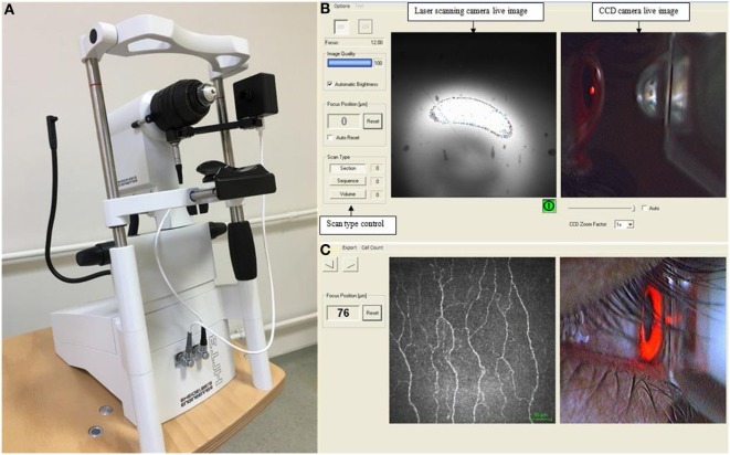
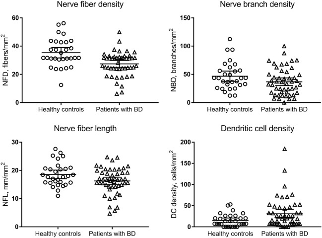
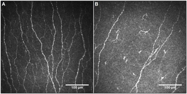
Central and peripheral nervous system involvement may occur during the course of Behçet's disease (BD). corneal confocal microscopy (CCM) can detect corneal small fiber damage and immune cell density. The aim of this study was to assess central corneal sensitivity, corneal subepithelial nerve plexus morphology and dendritic cell (DC) density in patients with BD. Forty-nine consecutive patients with BD and 30 healthy control subjects were included in this cross-sectional study conducted at a tertiary referral university hospital. Central corneal sensitivity was measured using the contact corneal esthesiometer (Cochet-Bonnet; Luneau, France). The laser scanning CCM (Heidelberg, Germany) was used to quantify corneal nerve fiber density (NFD), nerve branch density (NBD), nerve fiber length (NFL), and DC density. There was a significant reduction in NFD ( = 0.001) and NFL ( = 0.031) and an increase in DC density ( = 0.038) in patients with BD compared to healthy controls, whereas corneal sensitivity ( = 0.066) and NBD ( = 0.067) did not differ significantly. There was no difference in corneal sensitivity, corneal nerve parameters, or DC density between BD patients with [ = 18 (36.7%)] and without a previous history of uveitis ( > 0.05 for all). Disease duration [median (IQR), 6.5 (4.0-14.5) years] correlated with corneal sensitivity (ρ = -0.463;  = 0.001) and NFD (ρ = -0.304;  = 0.034) and corneal sensitivity correlated with NFD (ρ = 0.411;  = 0.003) and NFL (ρ = 0.295;  = 0.039) in patients with BD. CCM demonstrates corneal sub-basal nerve fiber loss and increased DC density, providing a non-invasive ophthalmic means to identify peripheral neuropathy and inflammation in patients with BD.

摘要

白塞病(BD)病程中可能会出现中枢和周围神经系统受累。角膜共焦显微镜检查(CCM)可检测角膜小纤维损伤和免疫细胞密度。本研究旨在评估BD患者的中央角膜敏感性、角膜上皮下神经丛形态和树突状细胞(DC)密度。在一所三级转诊大学医院进行的这项横断面研究中,纳入了49例连续的BD患者和30名健康对照者。使用接触式角膜感觉计(Cochet-Bonnet;法国吕诺)测量中央角膜敏感性。使用激光扫描CCM(德国海德堡)量化角膜神经纤维密度(NFD)、神经分支密度(NBD)、神经纤维长度(NFL)和DC密度。与健康对照相比,BD患者的NFD(P = 0.001)和NFL(P = 0.031)显著降低,DC密度增加(P = 0.038),而角膜敏感性(P = 0.066)和NBD(P = 0.067)无显著差异。有葡萄膜炎既往史的BD患者[18例(36.7%)]与无葡萄膜炎既往史的BD患者在角膜敏感性、角膜神经参数或DC密度方面无差异(所有P均>0.05)。病程[中位数(四分位间距),6.5(4.0 - 14.5)年]与BD患者的角膜敏感性(ρ = -0.463;P = 0.001)和NFD(ρ = -0.304;P = 0.034)相关,且角膜敏感性与BD患者的NFD(ρ = 0.411;P = 0.003)和NFL(ρ = 0.295;P = 0.039)相关。CCM显示角膜基底膜下神经纤维丢失和DC密度增加,为识别BD患者的周围神经病变和炎症提供了一种非侵入性眼科手段。

https://cdn.ncbi.nlm.nih.gov/pmc/blobs/7daa/5882821/9f17946d986a/fneur-09-00204-g003.jpg
https://cdn.ncbi.nlm.nih.gov/pmc/blobs/7daa/5882821/77f946d8fa71/fneur-09-00204-g001.jpg
https://cdn.ncbi.nlm.nih.gov/pmc/blobs/7daa/5882821/ccb8df2e628f/fneur-09-00204-g002.jpg
https://cdn.ncbi.nlm.nih.gov/pmc/blobs/7daa/5882821/9f17946d986a/fneur-09-00204-g003.jpg
https://cdn.ncbi.nlm.nih.gov/pmc/blobs/7daa/5882821/77f946d8fa71/fneur-09-00204-g001.jpg
https://cdn.ncbi.nlm.nih.gov/pmc/blobs/7daa/5882821/ccb8df2e628f/fneur-09-00204-g002.jpg
https://cdn.ncbi.nlm.nih.gov/pmc/blobs/7daa/5882821/9f17946d986a/fneur-09-00204-g003.jpg

相似文献

1
Confocal Microscopic Evaluation of Corneal Nerve Fibers and Dendritic Cells in Patients With Behçet's Disease.白塞病患者角膜神经纤维和树突状细胞的共聚焦显微镜评估
Front Neurol. 2018 Mar 28;9:204. doi: 10.3389/fneur.2018.00204. eCollection 2018.
2
Corneal confocal microscopy detects corneal nerve damage and increased dendritic cells in Fabry disease.角膜共焦显微镜检测法可发现法布里病的角膜神经损伤和树突状细胞增多。
Sci Rep. 2018 Aug 16;8(1):12244. doi: 10.1038/s41598-018-30688-z.
3
Use of Corneal Confocal Microscopy to Detect Corneal Nerve Loss and Increased Dendritic Cells in Patients With Multiple Sclerosis.使用角膜共焦显微镜检测多发性硬化症患者的角膜神经损伤和树突状细胞增多
JAMA Ophthalmol. 2017 Jul 1;135(7):777-782. doi: 10.1001/jamaophthalmol.2017.1590.
4
Subclinical Corneal Nerve Fiber Damage and Immune Cell Activation in Systemic Lupus Erythematosus: A Corneal Confocal Microscopy Study.系统性红斑狼疮患者亚临床角膜神经纤维损伤和免疫细胞激活:一项角膜共聚焦显微镜研究。
Transl Vis Sci Technol. 2021 Dec 1;10(14):10. doi: 10.1167/tvst.10.14.10.
5
Corneal innervation changes ın Alzheimer's: implications for sensory dysfunction.阿尔茨海默病患者的角膜神经支配变化:对感觉功能障碍的影响。
Int Ophthalmol. 2024 Jun 24;44(1):270. doi: 10.1007/s10792-024-03162-1.
6
[Early diagnosis of painful diabetic neuropathy by corneal confocal microscopy].[通过角膜共焦显微镜早期诊断疼痛性糖尿病神经病变]
Zhonghua Yi Xue Za Zhi. 2014 Sep 9;94(33):2602-6.
7
Small-Fiber Neuropathy Is Associated With Corneal Nerve and Dendritic Cell Alterations: An In Vivo Confocal Microscopy Study.小纤维神经病变与角膜神经及树突状细胞改变相关:一项共聚焦显微镜活体研究
Cornea. 2015 Sep;34(9):1114-9. doi: 10.1097/ICO.0000000000000535.
8
[The value of corneal confocal microscopy in diagnosing early small fibre neuropathy].[角膜共焦显微镜在早期小纤维神经病变诊断中的价值]
Zhonghua Nei Ke Za Zhi. 2014 Oct;53(10):778-82.
9
[Corneal confocal microscopy detects small-fiber neuropathy in patients with amyotrophic lateral sclerosis].[角膜共焦显微镜检测肌萎缩侧索硬化症患者的小纤维神经病变]
Zhonghua Nei Ke Za Zhi. 2022 Jan 1;61(1):77-81. doi: 10.3760/cma.j.cn112138-20210207-00114.
10
In vivo confocal microscopy detects bilateral changes of corneal immune cells and nerves in unilateral herpes zoster ophthalmicus.在体共聚焦显微镜检测单侧带状疱疹性眼病双侧角膜免疫细胞和神经的变化。
Ocul Surf. 2018 Jan;16(1):101-111. doi: 10.1016/j.jtos.2017.09.004. Epub 2017 Sep 18.

引用本文的文献

1
Decoding Behcet's Uveitis: an In-depth review of pathogenesis and therapeutic advances.贝赫切特葡萄膜炎的解码:发病机制和治疗进展的深入综述。
J Neuroinflammation. 2024 May 22;21(1):133. doi: 10.1186/s12974-024-03123-6.
2
Corneal nerve loss and increased Langerhans cells are associated with disease severity in patients with rheumatoid arthritis.角膜神经丢失和朗格汉斯细胞增多与类风湿关节炎患者的疾病严重程度相关。
Eye (Lond). 2023 Oct;37(14):2950-2955. doi: 10.1038/s41433-023-02447-6. Epub 2023 Feb 18.
3
Corneal Sub-Basal Nerve Plexus in Non-Diabetic Small Fiber Polyneuropathies and the Diagnostic Role of In Vivo Corneal Confocal Microscopy.

本文引用的文献

1
Clinical and imaging features of spinal cord type of neuro Behçet disease: A case report and systematic review.神经白塞病脊髓型的临床及影像学特征:一例病例报告及系统综述
Medicine (Baltimore). 2017 Oct;96(40):e7958. doi: 10.1097/MD.0000000000007958.
2
CIDP and other inflammatory neuropathies in diabetes - diagnosis and management.糖尿病相关 CIDP 和其他炎性神经病——诊断与管理。
Nat Rev Neurol. 2017 Oct;13(10):599-611. doi: 10.1038/nrneurol.2017.123. Epub 2017 Sep 15.
3
Corneal confocal microscopy is a rapid reproducible ophthalmic technique for quantifying corneal nerve abnormalities.
非糖尿病性小纤维多发性神经病中的角膜基底膜下神经丛及活体共聚焦显微镜的诊断作用
J Clin Med. 2023 Jan 13;12(2):664. doi: 10.3390/jcm12020664.
4
Artificial Intelligence and Corneal Confocal Microscopy: The Start of a Beautiful Relationship.人工智能与角膜共焦显微镜:美好关系的开端。
J Clin Med. 2022 Oct 20;11(20):6199. doi: 10.3390/jcm11206199.
5
Investigation of anterior segment structures of the eye in Behçet's disease using in vivo confocal microscopy.应用活体共聚焦显微镜研究 Behçet 病眼前段结构。
Graefes Arch Clin Exp Ophthalmol. 2022 Dec;260(12):3897-3902. doi: 10.1007/s00417-022-05846-9. Epub 2022 Sep 30.
6
In vivo confocal microscopy for eyes with Behçet's disease: a missing piece of the puzzle.白塞病眼部的活体共聚焦显微镜检查:难题中的缺失环节。
Graefes Arch Clin Exp Ophthalmol. 2022 Dec;260(12):3779-3780. doi: 10.1007/s00417-022-05848-7. Epub 2022 Sep 29.
7
Changes in corneal nerve morphology and function in patients with dry eyes having type 2 diabetes.2型糖尿病干眼症患者角膜神经形态和功能的变化
World J Clin Cases. 2022 Apr 6;10(10):3014-3026. doi: 10.12998/wjcc.v10.i10.3014.
8
Corneal Confocal Microscopy for Assessment of Non-Neurological Autoimmune Diseases: A Meta-Analysis.用于评估非神经自身免疫性疾病的角膜共焦显微镜检查:一项荟萃分析。
Front Med (Lausanne). 2022 Mar 9;9:809164. doi: 10.3389/fmed.2022.809164. eCollection 2022.
9
Corneal Confocal Microscopy to Image Small Nerve Fiber Degeneration: Ophthalmology Meets Neurology.角膜共焦显微镜成像观察小神经纤维退变:眼科与神经科的交汇
Front Pain Res (Lausanne). 2021 Aug 19;2:725363. doi: 10.3389/fpain.2021.725363. eCollection 2021.
10
The impact of sensory neuropathy and inflammation on epithelial wound healing in diabetic corneas.感觉神经病变和炎症对糖尿病角膜上皮伤口愈合的影响。
Prog Retin Eye Res. 2022 Jul;89:101039. doi: 10.1016/j.preteyeres.2021.101039. Epub 2022 Jan 4.
角膜共焦显微镜检查是一种用于量化角膜神经异常的快速可重复的眼科技术。
PLoS One. 2017 Aug 17;12(8):e0183040. doi: 10.1371/journal.pone.0183040. eCollection 2017.
4
Corneal Confocal Microscopy: An Imaging Endpoint for Axonal Degeneration in Multiple Sclerosis.角膜共焦显微镜检查:多发性硬化症轴突退变的一种成像终点指标
Invest Ophthalmol Vis Sci. 2017 Jul 1;58(9):3677-3681. doi: 10.1167/iovs.17-22050.
5
Diagnostic utility of corneal confocal microscopy and intra-epidermal nerve fibre density in diabetic neuropathy.角膜共焦显微镜检查和表皮内神经纤维密度在糖尿病神经病变中的诊断效用
PLoS One. 2017 Jul 18;12(7):e0180175. doi: 10.1371/journal.pone.0180175. eCollection 2017.
6
Comparison of optical coherence tomographic findings between Behcet disease patients with and without ocular involvement and healthy subjects.有眼部受累和无眼部受累的白塞病患者与健康受试者之间光学相干断层扫描结果的比较。
Arq Bras Oftalmol. 2017 Mar-Apr;80(2):69-73. doi: 10.5935/0004-2749.20170018.
7
Use of Corneal Confocal Microscopy to Detect Corneal Nerve Loss and Increased Dendritic Cells in Patients With Multiple Sclerosis.使用角膜共焦显微镜检测多发性硬化症患者的角膜神经损伤和树突状细胞增多
JAMA Ophthalmol. 2017 Jul 1;135(7):777-782. doi: 10.1001/jamaophthalmol.2017.1590.
8
Macular Inner Retinal Layer Thickening and Outer Retinal Layer Damage Correlate With Visual Acuity During Remission in Behcet's Disease.白塞病缓解期黄斑视网膜内层增厚和外层视网膜损伤与视力相关。
Invest Ophthalmol Vis Sci. 2016 Oct 1;57(13):5470-5478. doi: 10.1167/iovs.16-19568.
9
Inflammatory optic neuropathy in Behçet's disease.白塞病中的炎性视神经病变。
Reumatismo. 2015 Dec 23;67(4):156-60. doi: 10.4081/reumatismo.2015.835.
10
Corneal confocal microscopy in chronic inflammatory demyelinating polyneuropathy.慢性炎症性脱髓鞘性多发性神经病的角膜共焦显微镜检查。
Ann Clin Transl Neurol. 2015 Dec 28;3(2):88-100. doi: 10.1002/acn3.275. eCollection 2016 Feb.