Ocular Surface Imaging Center and Cornea & Refractive Surgery Service, Massachusetts Eye & Ear Infirmary, Department of Ophthalmology, Harvard Medical School, 243 Charles Street, Boston, MA 02114, USA; Post-Graduate Program, Surgery Department, Pernambuco Federal University (UFPE), Recife, PE, Brazil.
Ocular Surface Imaging Center and Cornea & Refractive Surgery Service, Massachusetts Eye & Ear Infirmary, Department of Ophthalmology, Harvard Medical School, 243 Charles Street, Boston, MA 02114, USA.
Ocul Surf. 2018 Jan;16(1):101-111. doi: 10.1016/j.jtos.2017.09.004. Epub 2017 Sep 18.
To analyze bilateral corneal immune cell and nerve alterations in patients with unilateral herpes zoster ophthalmicus (HZO) by laser in vivo confocal microscopy (IVCM) and their correlation with corneal sensation and clinical findings.
This is a prospective, cross-sectional, controlled, single-center study. Twenty-four eyes of 24 HZO patients and their contralateral clinically unaffected eyes and normal controls (n = 24) were included. Laser IVCM (Heidelberg Retina Tomograph/Rostock Cornea Module), corneal esthesiometry (Cochet-Bonnet) were performed. Changes in corneal dendritiform cell (DC) density and morphology, number and length of subbasal nerve fibers and their correlation to corneal sensation, pain, lesion location, disease duration, and number of episodes were analyzed.
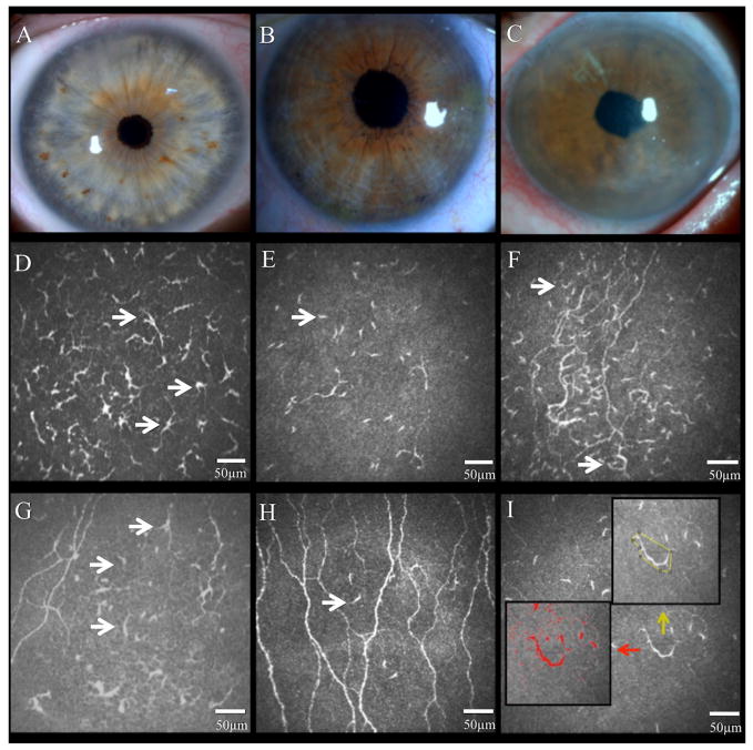
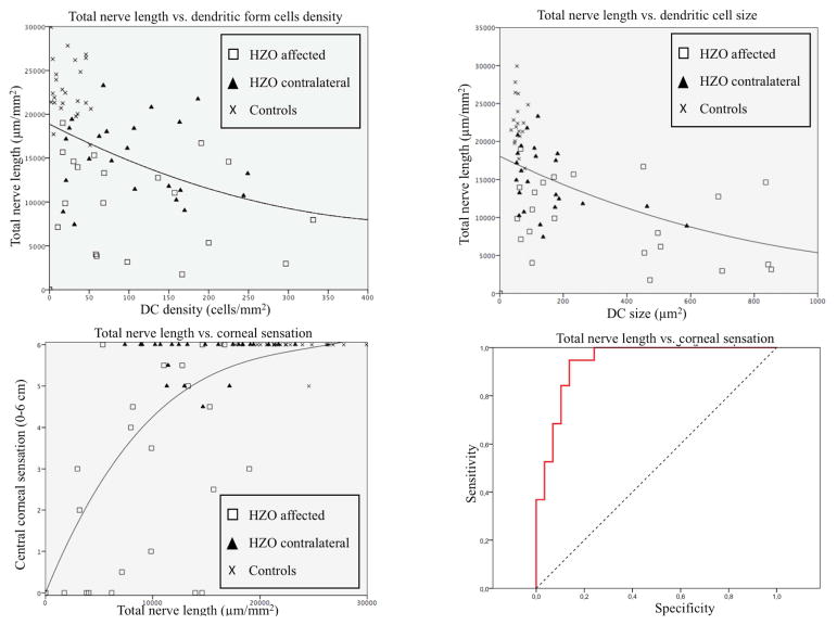
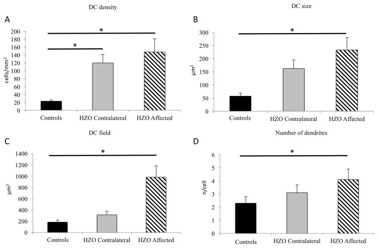
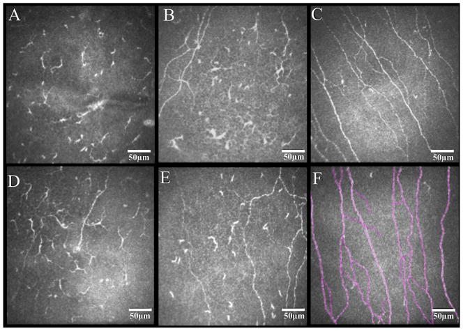
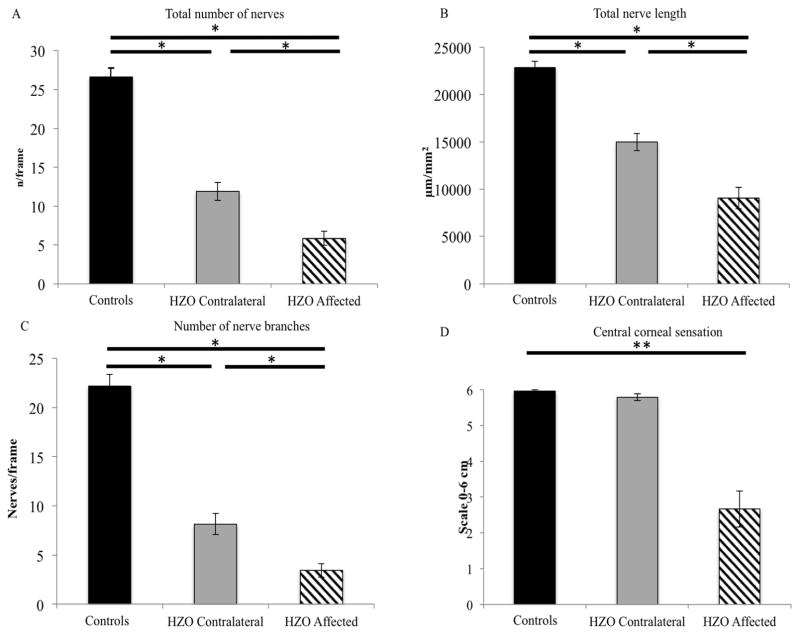
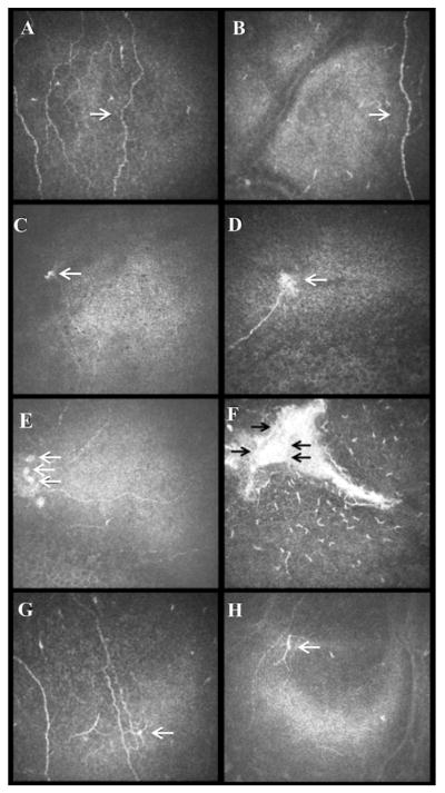
HZO-affected and contralateral eyes showed a significant increase in DC influx of the central cornea as compared to controls (147.4 ± 33.9, 120.1 ± 21.2, and 23.0 ± 3.6 cells/mm2; p < 0.0001). In HZO eyes DCs were larger in area (319.4 ± 59.8 μm; p < 0.001) and number of dendrites (3.5 ± 0.4 n/cell; p = 0.01) as compared to controls (52.2 ± 11.7, and 2.3 ± 0.5). DC density and size showed moderate negative correlation with total nerve length (R = -0.43 and R = -0.57, respectively; all p < 0.001). A higher frequency of nerve beading and activated DCs close to nerve fibers were detected specifically in pain patients.
Chronic unilateral HZO causes significant bilateral increase in corneal DC density and decrease of the corneal subbasal nerves as compared to controls. Negative correlation was observed for DC density and size to nerve parameters, suggesting interplay between the immune and nervous systems. Patients with chronic pain also showed increased nerve beading and activated DCs.
通过激光共聚焦显微镜(IVCM)分析单侧带状疱疹性眼病(HZO)患者双侧角膜免疫细胞和神经的变化,并分析其与角膜感觉和临床发现的相关性。
这是一项前瞻性、横断面、对照、单中心研究。纳入 24 例 HZO 患者的 24 只眼及其对侧临床无影响的眼和正常对照组(n=24)。行激光共聚焦显微镜(海德堡视网膜断层扫描仪/罗托卡角膜模块)和角膜知觉仪(Cochet-Bonnet)检查。分析角膜树突状细胞(DC)密度和形态变化、亚基底神经纤维数量和长度及其与角膜感觉、疼痛、病变部位、疾病持续时间和发作次数的相关性。
与对照组相比,HZO 受累眼和对侧眼的中央角膜 DC 内流显著增加(147.4±33.9、120.1±21.2 和 23.0±3.6 个细胞/mm2;p<0.0001)。HZO 眼的 DC 面积(319.4±59.8μm;p<0.001)和树突数(3.5±0.4 个/细胞;p=0.01)均大于对照组(52.2±11.7 和 2.3±0.5)。DC 密度和大小与总神经长度呈中度负相关(R=-0.43 和 R=-0.57,均 p<0.001)。在疼痛患者中,特别是在疼痛患者中,检测到神经珠和靠近神经纤维的激活 DC 的频率更高。
与对照组相比,慢性单侧 HZO 导致双侧角膜 DC 密度显著增加,角膜亚基底神经减少。DC 密度和大小与神经参数呈负相关,提示免疫系统和神经系统之间存在相互作用。慢性疼痛患者也表现出神经珠和激活的 DC 增加。