Shen Yiming, Han Seong Kyu, Ryu Pan Dong
Department of Veterinary Pharmacology, College of Veterinary Medicine and Research Institute of Veterinary Science, Seoul National University, Seoul 08826, Korea.
Department of Oral Physiology, School of Dentistry and Institute of Oral Bioscience, Chonbuk National University, Jeonju 54896, Korea.
J Vet Sci. 2018 Jul 31;19(4):483-491. doi: 10.4142/jvs.2018.19.4.483.
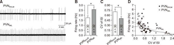
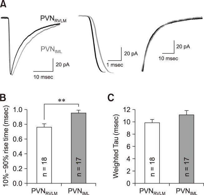
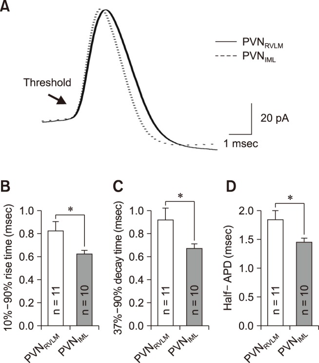
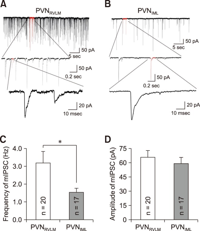
The hypothalamic paraventricular nucleus (PVN) contains two types of neurons projecting to either the rostral ventrolateral medulla (PVN) or the intermediolateral horn (IML) of the spinal cord (PVN). These two neuron groups are intermingled in the same subdivisions of the PVN and differentially regulate sympathetic outflow. However, electrophysiological evidence supporting such functional differences is largely lacking. Herein, we compared the electrophysiological properties of these neurons by using patch-clamp and retrograde-tracing techniques. Most neurons (>70%) in both groups spontaneously fired in the cell-attached mode. When compared to the PVN neurons, the PVN neurons had a lower firing rate and a more irregular firing pattern ( < 0.05). The PVN neurons showed smaller resting membrane potential, slower rise and decay times, and greater duration of spontaneous action potentials ( < 0.05). The PVN neurons received greater inhibitory synaptic inputs (frequency, < 0.05) with a shorter rise time ( < 0.05). Taken together, the results indicate that the two pre-sympathetic neurons differ in their intrinsic and extrinsic electrophysiological properties, which may explain the lower firing activity of the PVN neurons. The greater inhibitory synaptic inputs to the PVN neurons also imply that these neurons have more integrative roles in regulation of sympathetic activity.
下丘脑室旁核(PVN)包含两类神经元,它们分别投射至延髓头端腹外侧(RVLM)或脊髓中间外侧角(IML)(PVN)。这两组神经元在PVN的相同亚区相互交织,并对交感神经输出进行差异性调节。然而,很大程度上缺乏支持这种功能差异的电生理证据。在此,我们运用膜片钳和逆行追踪技术比较了这些神经元的电生理特性。两组中的大多数神经元(>70%)在细胞贴附模式下自发放电。与投射至RVLM的PVN神经元相比,投射至IML的PVN神经元放电频率更低且放电模式更不规则(P<0.05)。投射至IML的PVN神经元静息膜电位更小,上升和衰减时间更慢,自发动作电位持续时间更长(P<0.05)。投射至IML的PVN神经元接受更强的抑制性突触输入(频率,P<0.05),且上升时间更短(P<0.05)。综上所述,结果表明这两类交感节前神经元在内在和外在电生理特性方面存在差异,这可能解释了投射至IML的PVN神经元较低的放电活动。投射至IML的PVN神经元接受更强的抑制性突触输入也意味着这些神经元在交感神经活动调节中具有更多的整合作用。