Research Laboratory of Burns and Trauma, the 181st Hospital of Chinese PLA, Guilin, 541002, People's Republic of China.
Department of Burns, Plastic and Wound repair surgery, the 181st Hospital of Chinese PLA, Guilin, 541002, People's Republic of China.
Stem Cell Res Ther. 2018 Apr 12;9(1):101. doi: 10.1186/s13287-018-0856-7.
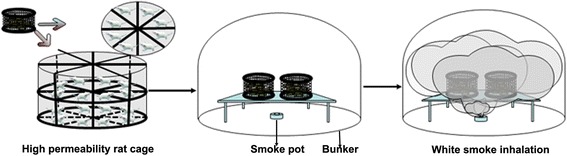
White smoke inhalation (WSI) is an uncommon but potentially deadly cause of acute lung injury and acute respiratory distress syndrome for which no effective pharmaceutical treatment has been developed. This study aimed to determine the protective effects of human amnion-derived mesenchymal stem cells (hAMSCs) against WSI-induced lung injury in rats.
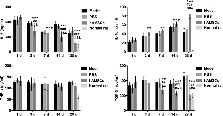
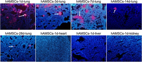
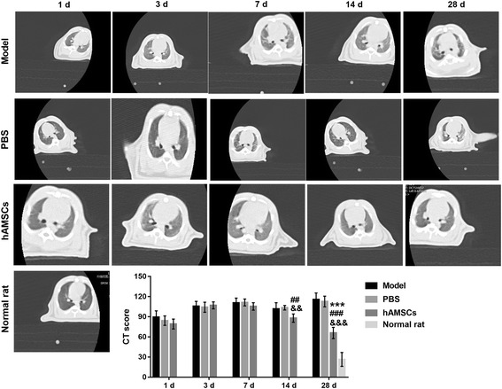
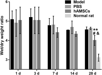
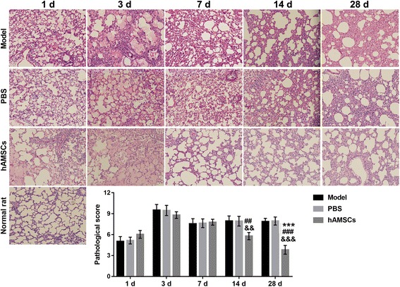
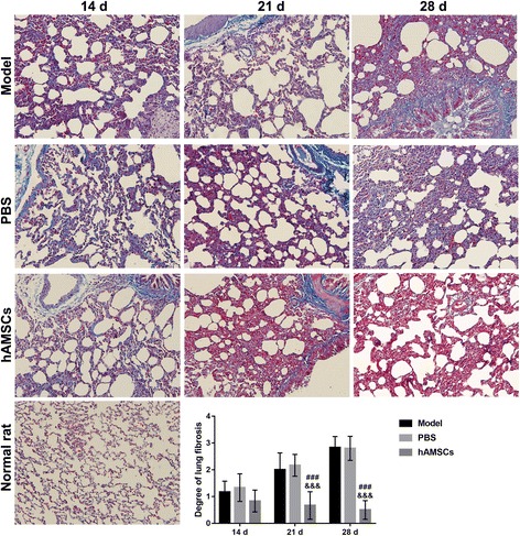
hAMSCs were injected into rats via the tail vein 4 h after WSI. At 1, 3, 7, 14, and 28 days after cell injection, hAMSCs labeled with PKH26 in lung, heart, liver, and kidney tissues were observed by fluorescence microscopy. The lung injury score was determined by hematoxylin and eosin staining. Lung fibrosis was assessed by Masson's trichrome staining. The computed tomography (CT) score was assessed by CT scanning. The wet/dry weight ratio was calculated. The levels of interleukin (IL)-1β, IL-6, and IL-10 were determined by enzyme-linked immunosorbent assays. The expression of surfactant protein (SP)-A, SP-C, and SP-D was measured by Western blotting.
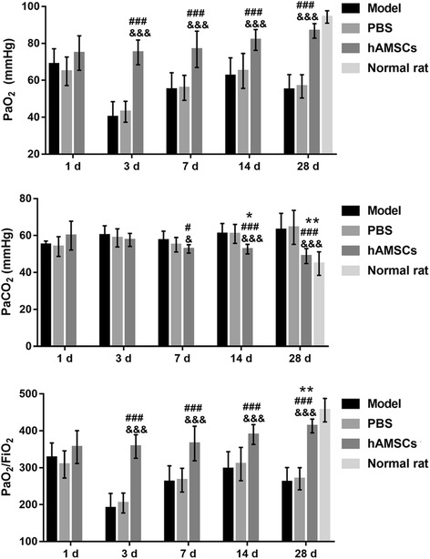
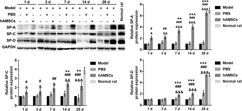
The injected hAMSCs were primarily distributed in the lung tissues in WSI-induced rats. Compared with the model and phosphate-buffered saline (PBS) group, hAMSC treatment led to reduced lung injury, lung fibrosis, CT score, and inflammation levels in WSI-induced mice. hAMSC treatment also resulted in increased cell retention in the lung, partial pressure of oxygen (PaO), and PaO/fraction of inspired oxygen (FiO) levels, and pulmonary SP-A, SP-C, and SP-D expression compared with that in the model and PBS group.
hAMSCs are a potential cell-based therapy for WSI-induced lung injury.
白烟吸入(WSI)是一种罕见但潜在致命的急性肺损伤和急性呼吸窘迫综合征的原因,目前尚未开发出有效的药物治疗方法。本研究旨在确定人羊膜来源间充质干细胞(hAMSCs)对 WSI 诱导的大鼠肺损伤的保护作用。
WSI 后 4 小时通过尾静脉向大鼠注射 hAMSCs。在细胞注射后 1、3、7、14 和 28 天,通过荧光显微镜观察 PKH26 标记的肺、心、肝和肾组织中的 hAMSCs。通过苏木精和伊红染色确定肺损伤评分。通过 Masson 三色染色评估肺纤维化。通过 CT 扫描评估 CT 评分。计算湿/干重比。通过酶联免疫吸附试验测定白细胞介素(IL)-1β、IL-6 和 IL-10 的水平。通过 Western 印迹测定表面活性蛋白(SP)-A、SP-C 和 SP-D 的表达。
注射的 hAMSCs 主要分布在 WSI 诱导的大鼠肺组织中。与模型和磷酸盐缓冲盐水(PBS)组相比,hAMSC 治疗导致 WSI 诱导的小鼠肺损伤、肺纤维化、CT 评分和炎症水平降低。与模型和 PBS 组相比,hAMSC 治疗还导致肺细胞保留增加、氧分压(PaO)和 PaO/吸入氧分数(FiO)水平升高,以及肺 SP-A、SP-C 和 SP-D 表达增加。
hAMSCs 是一种潜在的细胞治疗方法,可用于治疗 WSI 诱导的肺损伤。