Zlotorowicz M, Czubak-Wrzosek M, Wrzosek P, Czubak J
Centre of Postgraduate Medical Education, Warsaw, Poland.
Department of Orthopaedics, Pediatric Orthopaedics and Traumatology, Gruca Teaching Hospital, Otwock, Poland.
Surg Radiol Anat. 2018 May;40(5):515-520. doi: 10.1007/s00276-018-2012-6. Epub 2018 Apr 12.
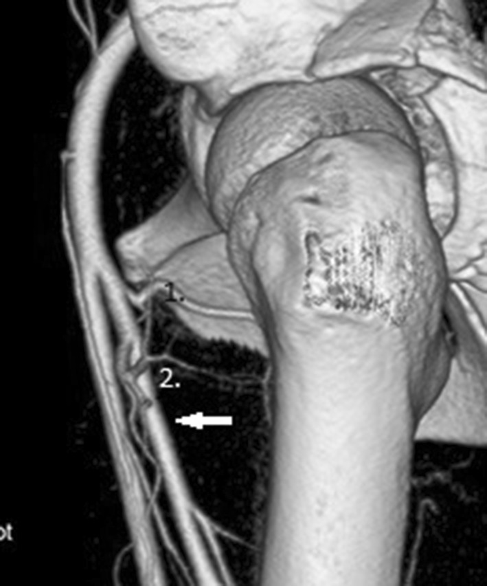
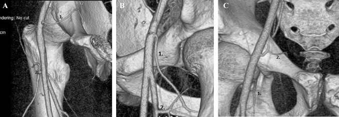
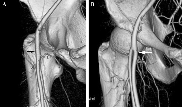
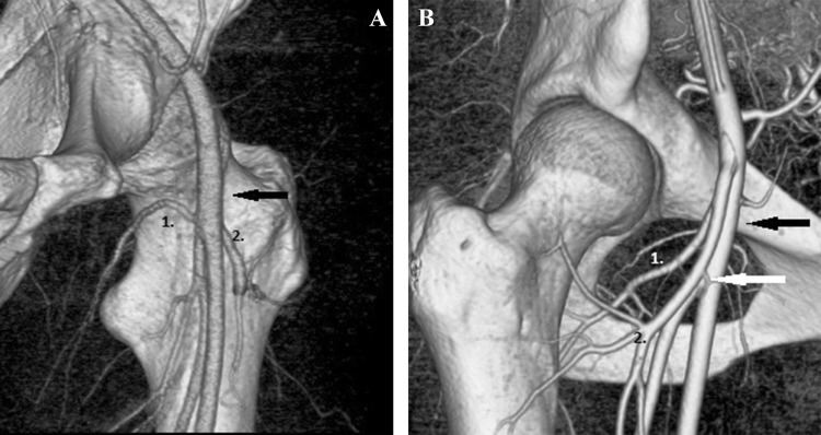
The most significant blood vessel supplying the hip joint is the medial femoral circumflex artery (MFCA). MFCA with lateral femoral circumflex artery (LFCA) are the first branches of the deep femoral artery (DFA) or they originate directly from the common femoral artery (CFA) or superficial femoral artery (SFA).
We analyzed 100 CT angiogram of the hip region [72 men, 28 women; mean age 46.4 (14-80)] to assess the frequency of each type of division of the MFCA and LFCA from either the DFA or directly from the CFA or SFA. To assess the variations on each side in one individual we analyzed both hips in 73 patients [mean age 46.6 (14-80)].
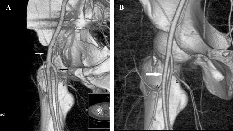
Many different types of division have been described. The most frequent one in which both the MFCA and LFCA originate from the DFA, was observed in 50% of patients. In 31% of hips the MFCA originates from the CFA. In our study, a normal origin of the obturator artery from the internal iliac artery was observed in 67% of patients and an atypical origin, called corona mortis was observed in 33% of patients.
The deep branch of the MFCA is the main artery supplying the femoral head, it is at risk during surgical approach to the hip joint. The atypical anastomosis called corona mortis is also at risk while performing the approach to pubic bone. Therefore, knowledge of their topography is very important.
供应髋关节的最重要血管是股内侧旋股动脉(MFCA)。MFCA与股外侧旋股动脉(LFCA)是股深动脉(DFA)的第一分支,或者它们直接起源于股总动脉(CFA)或股浅动脉(SFA)。
我们分析了100例髋关节区域的CT血管造影[72例男性,28例女性;平均年龄46.4(14 - 80岁)],以评估MFCA和LFCA从DFA或直接从CFA或SFA分出的每种类型的频率。为了评估个体两侧的变异情况,我们分析了73例患者[平均年龄46.6(14 - 80岁)]的双侧髋关节。
已描述了许多不同类型的分支。最常见的情况是MFCA和LFCA均起源于DFA,在50%的患者中观察到。在31%的髋关节中,MFCA起源于CFA。在我们的研究中,67%的患者闭孔动脉正常起源于髂内动脉,33%的患者观察到一种非典型起源,称为死亡冠。
MFCA的深支是供应股骨头的主要动脉,在髋关节手术入路过程中存在风险。在耻骨手术入路时,称为死亡冠的非典型吻合也有风险。因此,了解它们的解剖位置非常重要。