文献检索文档翻译深度研究
Suppr Zotero 插件Zotero 插件
邀请有礼套餐&价格历史记录

新学期,新优惠

限时优惠:9月1日-9月22日

30天高级会员仅需29元

1天体验卡首发特惠仅需5.99元

了解详情
不再提醒
插件&应用
Suppr Zotero 插件Zotero 插件浏览器插件Mac 客户端Windows 客户端微信小程序
高级版
套餐订阅购买积分包
AI 工具
文献检索文档翻译深度研究
关于我们
关于 Suppr公司介绍联系我们用户协议隐私条款
关注我们

Suppr 超能文献

核心技术专利:CN118964589B侵权必究
粤ICP备2023148730 号-1Suppr @ 2025

微观结构磁共振成像能否检测无症状性颈脊髓压迫患者的亚临床组织损伤?一项前瞻性队列研究。

Can microstructural MRI detect subclinical tissue injury in subjects with asymptomatic cervical spinal cord compression? A prospective cohort study.

作者信息

Martin Allan R, De Leener Benjamin, Cohen-Adad Julien, Cadotte David W, Nouri Aria, Wilson Jefferson R, Tetreault Lindsay, Crawley Adrian P, Mikulis David J, Ginsberg Howard, Fehlings Michael G

机构信息

Division of Neurosurgery, Department of Surgery, University of Toronto, Toronto, Ontario, Canada.

Institute of Biomedical Engineering, École Polytechnique de Montréal, Montréal, Québec, Canada.

出版信息

BMJ Open. 2018 Apr 13;8(4):e019809. doi: 10.1136/bmjopen-2017-019809.


DOI:10.1136/bmjopen-2017-019809
PMID:29654015
原文链接:https://pmc.ncbi.nlm.nih.gov/articles/PMC5905727/
Abstract

OBJECTIVES: Degenerative cervical myelopathy (DCM) involves extrinsic spinal cord compression causing tissue injury and neurological dysfunction. Asymptomatic spinal cord compression (ASCC) is more common, but its significance is poorly defined. This study investigates if: (1) ASCC can be automatically diagnosed using spinal cord shape analysis; (2) multiparametric quantitative MRI can detect similar spinal cord tissue injury as previously observed in DCM. DESIGN: Prospective observational longitudinal cohort study. SETTING: Single centre, tertiary care and research institution. PARTICIPANTS: 40 neurologically intact subjects (19 female, 21 male) divided into groups with and without ASCC. INTERVENTIONS: None. OUTCOME MEASURES: 
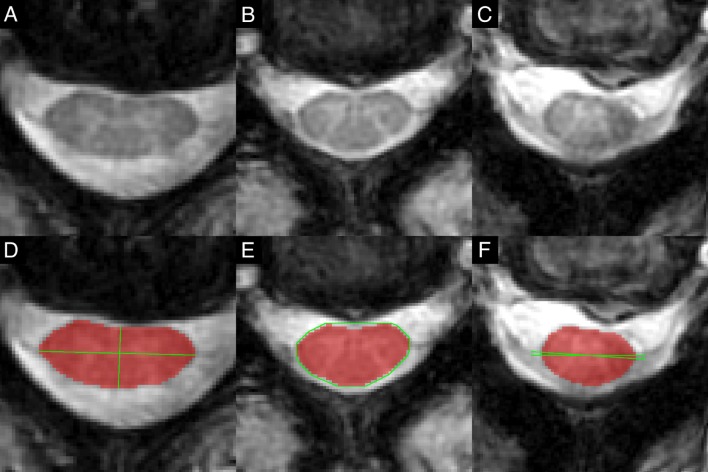
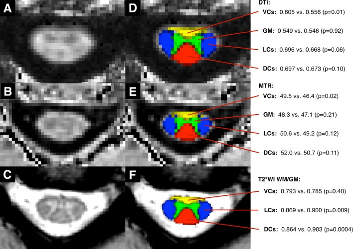
Clinical assessments: modified Japanese Orthopaedic Association score and physical examination. 3T MRI assessments: automated morphometric analysis compared with consensus ratings of spinal cord compression, and measures of tissue injury: cross-sectional area, diffusion fractional anisotropy, magnetisation transfer ratio and T2*-weighted imaging white to grey matter signal intensity ratio (T2*WI WM/GM) extracted from rostral (C1-3), caudal (C6-7) and maximally compressed levels. RESULTS: 
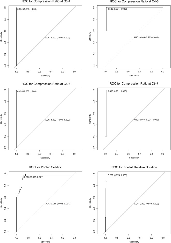
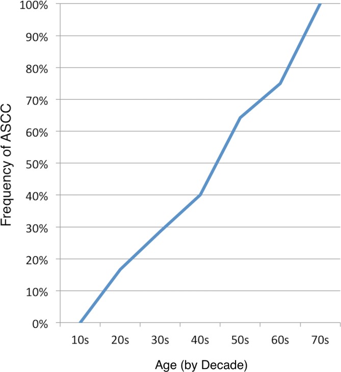
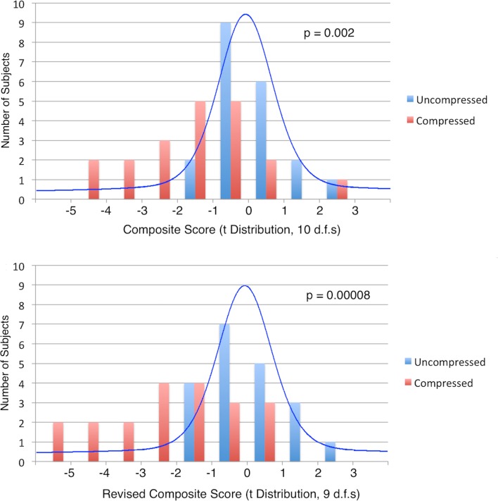
ASCC was present in 20/40 subjects. Diagnosis with automated shape analysis showed area under the curve >97%. Five MRI metrics showed differences suggestive of tissue injury in ASCC compared with uncompressed subjects (p<0.05), while a composite of all 10 measures (average of z scores) showed highly significant differences (p=0.002). At follow-up (median 21 months), two ASCC subjects developed DCM. CONCLUSIONS: ASCC appears to be common and can be accurately and objectively diagnosed with automated morphometric analysis. Quantitative MRI appears to detect subclinical tissue injury in ASCC prior to the onset of neurological symptoms and signs. These findings require further validation, but offer the intriguing possibility of presymptomatic diagnosis and treatment of DCM and other spinal pathologies.

摘要

目的:退行性颈椎脊髓病(DCM)涉及脊髓外部受压,导致组织损伤和神经功能障碍。无症状脊髓受压(ASCC)更为常见,但其意义尚不明确。本研究调查:(1)能否使用脊髓形态分析自动诊断ASCC;(2)多参数定量MRI能否检测到与先前在DCM中观察到的类似脊髓组织损伤。 设计:前瞻性观察性纵向队列研究。 地点:单中心三级医疗和研究机构。 参与者:40名神经功能正常的受试者(19名女性,21名男性),分为有ASCC组和无ASCC组。 干预措施:无。 观察指标:临床评估:改良日本骨科协会评分和体格检查。3T MRI评估:自动形态计量分析与脊髓受压的一致性评级比较,以及组织损伤测量:从延髓(C1 - 3)、尾端(C6 - 7)和最大受压水平提取的横截面积、扩散分数各向异性、磁化传递率和T2加权成像白质与灰质信号强度比(T2WI WM/GM)。 结果:40名受试者中有20名存在ASCC。自动形状分析诊断的曲线下面积>97%。与未受压受试者相比,5项MRI指标显示ASCC中存在提示组织损伤的差异(p<0.05),而所有10项测量指标的综合指标(z评分平均值)显示出高度显著差异(p = 0.002)。随访(中位时间21个月)时,2名ASCC受试者发展为DCM。 结论:ASCC似乎很常见,可通过自动形态计量分析准确、客观地诊断。定量MRI似乎能在神经症状和体征出现之前检测到ASCC中的亚临床组织损伤。这些发现需要进一步验证,但为DCM和其他脊柱疾病的症状前诊断和治疗提供了有趣的可能性。

https://cdn.ncbi.nlm.nih.gov/pmc/blobs/a7be/5905727/1a323e2b04a0/bmjopen-2017-019809f05.jpg
https://cdn.ncbi.nlm.nih.gov/pmc/blobs/a7be/5905727/cd11d15a0f0a/bmjopen-2017-019809f01.jpg
https://cdn.ncbi.nlm.nih.gov/pmc/blobs/a7be/5905727/aad26b59e33c/bmjopen-2017-019809f02.jpg
https://cdn.ncbi.nlm.nih.gov/pmc/blobs/a7be/5905727/67c9f06ab528/bmjopen-2017-019809f03.jpg
https://cdn.ncbi.nlm.nih.gov/pmc/blobs/a7be/5905727/ebea1bbf0512/bmjopen-2017-019809f04.jpg
https://cdn.ncbi.nlm.nih.gov/pmc/blobs/a7be/5905727/1a323e2b04a0/bmjopen-2017-019809f05.jpg

相似文献

[1]
Can microstructural MRI detect subclinical tissue injury in subjects with asymptomatic cervical spinal cord compression? A prospective cohort study.

BMJ Open. 2018-4-13

[2]
A Novel MRI Biomarker of Spinal Cord White Matter Injury: T2*-Weighted White Matter to Gray Matter Signal Intensity Ratio.

AJNR Am J Neuroradiol. 2017-6

[3]
Early neurological changes in aging cervical spine: insights from PROMIS mobility assessment.

Geroscience. 2024-6

[4]
Correlation of magnetic resonance diffusion tensor imaging and clinical findings of cervical myelopathy.

Spine J. 2013-3-21

[5]
Is the type and/or co-existence of degenerative spinal pathology associated with the occurrence of degenerative cervical myelopathy? A single centre retrospective analysis of individuals with MRI defined cervical cord compression.

J Clin Neurosci. 2023-11

[6]
Clinically Feasible Microstructural MRI to Quantify Cervical Spinal Cord Tissue Injury Using DTI, MT, and T2*-Weighted Imaging: Assessment of Normative Data and Reliability.

AJNR Am J Neuroradiol. 2017-6

[7]
Tracking White and Gray Matter Degeneration along the Spinal Cord Axis in Degenerative Cervical Myelopathy.

J Neurotrauma. 2021-11-1

[8]
Reproducibility, temporal stability, and functional correlation of diffusion MR measurements within the spinal cord in patients with asymptomatic cervical stenosis or cervical myelopathy.

J Neurosurg Spine. 2018-5

[9]
Predictors of symptomatic myelopathy in degenerative cervical spinal cord compression.

Brain Behav. 2017-8-11

[10]
Diffusion magnetic resonance imaging reveals tract-specific microstructural correlates of electrophysiological impairments in non-myelopathic and myelopathic spinal cord compression.

Eur J Neurol. 2021-11

引用本文的文献

[1]
Quantitative assessment of asymptomatic spinal cord compression using MRI: a multi-center study.

Geroscience. 2025-8-15

[2]
Body size and intracranial volume interact with the structure of the central nervous system: A multi-center in vivo neuroimaging study.

Imaging Neurosci (Camb). 2025-5-7

[3]
A Systematic Review of Current Terminology for Conditions Preceding Degenerative Cervical Myelopathy: Evidence Synthesis to Inform an AO Spine Expert Opinion Statement.

Global Spine J. 2025-4-30

[4]
Cervical spinal cord morphometrics in degenerative cervical myelopathy: quantification using semi-automated normalized technique and correlation with neurological dysfunctions.

Spine J. 2024-11

[5]
Finite Element Analysis for Degenerative Cervical Myelopathy: Scoping Review of the Current Findings and Design Approaches, Including Recommendations on the Choice of Material Properties.

JMIR Biomed Eng. 2024-3-28

[6]
Evidence-based commentary on the diagnosis, management, and further research of degenerative cervical spinal cord compression in the absence of clinical symptoms of myelopathy.

Front Neurol. 2024-5-10

[7]
Mapping the Degenerative Cervical Myelopathy Research Landscape: Topic Modeling of the Literature.

Global Spine J. 2025-4

[8]
Body size interacts with the structure of the central nervous system: A multi-center in vivo neuroimaging study.

bioRxiv. 2024-5-1

[9]
Toward a personalized approach: the promising horizon of degenerative cervical myelopathy.

Int J Surg. 2024-8-1

[10]
Reproducible Spinal Cord Quantitative MRI Analysis with the Spinal Cord Toolbox.

Magn Reson Med Sci. 2024-7-1

本文引用的文献

[1]
Clinically Feasible Microstructural MRI to Quantify Cervical Spinal Cord Tissue Injury Using DTI, MT, and T2*-Weighted Imaging: Assessment of Normative Data and Reliability.

AJNR Am J Neuroradiol. 2017-6

[2]
A Novel MRI Biomarker of Spinal Cord White Matter Injury: T2*-Weighted White Matter to Gray Matter Signal Intensity Ratio.

AJNR Am J Neuroradiol. 2017-6

[3]
Improved Diagnosis of Cervical Spondylotic Myelopathy with Contact Heat Evoked Potentials.

J Neurotrauma. 2017-6-15

[4]
SCT: Spinal Cord Toolbox, an open-source software for processing spinal cord MRI data.

Neuroimage. 2017-1-15

[5]
Prevalence and Imaging Characteristics of Nonmyelopathic and Myelopathic Spondylotic Cervical Cord Compression.

Spine (Phila Pa 1976). 2016-12-15

[6]
Spinal Cord MR Diffusion Properties in Patients with Degenerative Cervical Cord Compression.

J Neuroimaging. 2017-1

[7]
Magnetic resonance imaging assessment of degenerative cervical myelopathy: a review of structural changes and measurement techniques.

Neurosurg Focus. 2016-6

[8]
Tract-specific and age-related variations of the spinal cord microstructure: a multi-parametric MRI study using diffusion tensor imaging (DTI) and inhomogeneous magnetization transfer (ihMT).

NMR Biomed. 2016-6

[9]
Translating state-of-the-art spinal cord MRI techniques to clinical use: A systematic review of clinical studies utilizing DTI, MT, MWF, MRS, and fMRI.

Neuroimage Clin. 2015-12-4

[10]
Correlation of force control with regional spinal DTI in patients with cervical spondylosis without signs of spinal cord injury on conventional MRI.

Eur Radiol. 2016-3

文献AI研究员

20分钟写一篇综述,助力文献阅读效率提升50倍

立即体验

用中文搜PubMed

大模型驱动的PubMed中文搜索引擎

马上搜索

推荐工具

医学文档翻译智能文献检索