Granger Andre, Bricoune Ornella, Rajnauth Tina, Kimball David, Kimball Heather, Tubbs R Shane, Loukas Marios
Department of Anatomical Sciences, St. George's University School of Medicine, Grenada, West Indies.
Icu/Anaesthetics, Eric Williams Medical Sciences Complex.
Cureus. 2018 Feb 12;10(2):e2185. doi: 10.7759/cureus.2185.
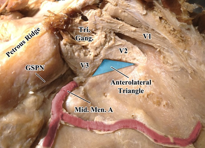
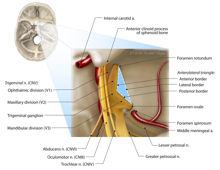
The anterolateral triangle is one of 10 surgical triangles of the cavernous sinus and serves as an important anatomic landmark for the skull base surgeon. There are few studies in the English literature that have precisely defined and measured the borders of the anterolateral triangle and little agreement has been made regarding the nomenclature within the English literature. A total of 12 midsagittally hemisected adult human cadaveric head halves were dissected to expose the anterolateral triangle. The triangle was defined and measurements of the anterior, posterior, and lateral borders were taken. The mean lengths and standard deviations of the anterior, posterior, and lateral borders were 8.3 ± 2.2 mm, 5.9 ± 2.0 mm, and 11.5 ± 2.9 mm, respectively. The mean area and standard deviation were 20.46 ± 9.30 mm. The anterolateral triangle is helpful in understanding and planning surgical approaches to the cavernous sinus and middle cranial fossa. As such, normal anatomic relationships and the sizes of the anterolateral triangle must first be recognized to better access the pathologic changes within and around this region.
前外侧三角是海绵窦的10个手术三角之一,是颅底外科医生重要的解剖标志。英文文献中很少有研究精确界定和测量前外侧三角的边界,对于英文文献中的命名也很少有一致意见。共解剖了12个经正中矢状面半切的成人尸体头部半侧,以暴露前外侧三角。对该三角进行了界定,并测量了其前、后和外侧边界。前、后和外侧边界的平均长度及标准差分别为8.3±2.2毫米、5.9±2.0毫米和11.5±2.9毫米。平均面积及标准差为20.46±9.30平方毫米。前外侧三角有助于理解和规划海绵窦及中颅窝的手术入路。因此,必须首先认识前外侧三角的正常解剖关系及大小,以便更好地处理该区域内及周围的病理变化。