Komatsu Fuminari, Oda Shinri, Shimoda Masami, Imai Masaaki, Shigematsu Hideaki, Komatsu Mika, Tschabitscher Manfred, Matsumae Mitsunori
Department of Neurosurgery, Tokai University Hachioji Hospital.
Neurol Med Chir (Tokyo). 2014;54(12):1004-8. doi: 10.2176/nmc.oa.2014-0092. Epub 2014 Nov 29.
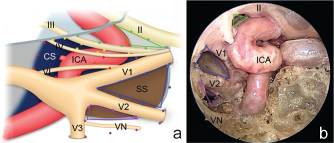
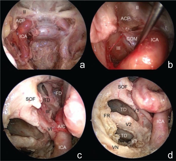
The lateral limit of endoscopic endonasal surgery has yet to be defined. The aim of this study was to investigate the lateral limit of endoscopic endonasal surgery at the level of the sphenoid sinus. Access from the sphenoid sinus to the middle cranial fossa through the cavernous sinus triangles was evaluated by cadaver dissection. Anatomical analysis demonstrated that the medial temporal dura mater was exposed through the anterior area of the clinoidal triangle, anteromedial triangle, and superior area of the anterolateral triangle, indicating potential corridors to the middle cranial fossa. This study suggests that the cavernous sinus triangles are applicable in selected cases to manage middle cranial fossa lesions by endoscopic endonasal surgery.
鼻内镜下鼻内手术的外侧界限尚未明确。本研究的目的是探讨蝶窦水平鼻内镜下鼻内手术的外侧界限。通过尸体解剖评估从蝶窦经海绵窦三角进入中颅窝的情况。解剖分析表明,颞叶内侧硬脑膜可通过床突三角的前部区域、前内侧三角以及前外侧三角的上部区域暴露,提示存在通向中颅窝的潜在通道。本研究表明,海绵窦三角在特定病例中可用于鼻内镜下鼻内手术治疗中颅窝病变。