da Silva Sampaio Luzia, Kubrusly Regina C C, Colli Yolanda P, Trindade Priscila P, Ribeiro-Resende Victor T, Einicker-Lamas Marcelo, Paes-de-Carvalho Roberto, Gardino Patricia F, de Mello Fernando G, De Melo Reis Ricardo A
Laboratório de Neuroquímica, Instituto de Biofísica Carlos Chagas Filho, Universidade Federal do Rio de Janeiro, Rio de Janeiro, Brazil.
Laboratório de Neurofarmacologia, Instituto Biomédico, Universidade Federal Fluminense, Niterói, Brazil.
Front Cell Neurosci. 2018 Mar 12;12:58. doi: 10.3389/fncel.2018.00058. eCollection 2018.
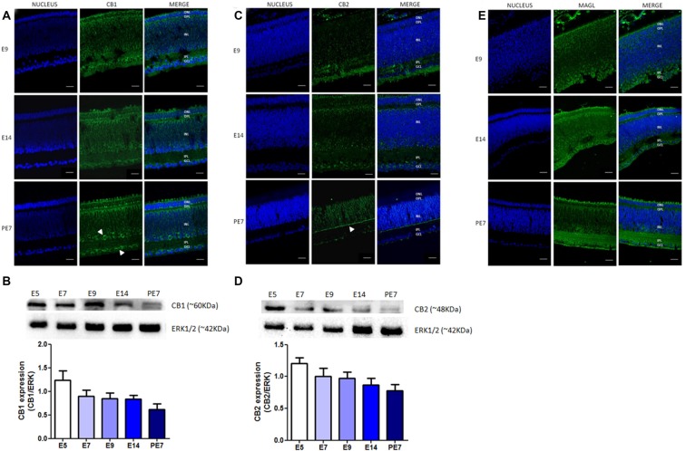
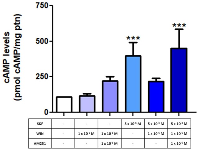
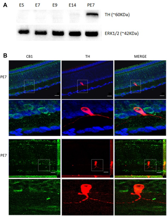
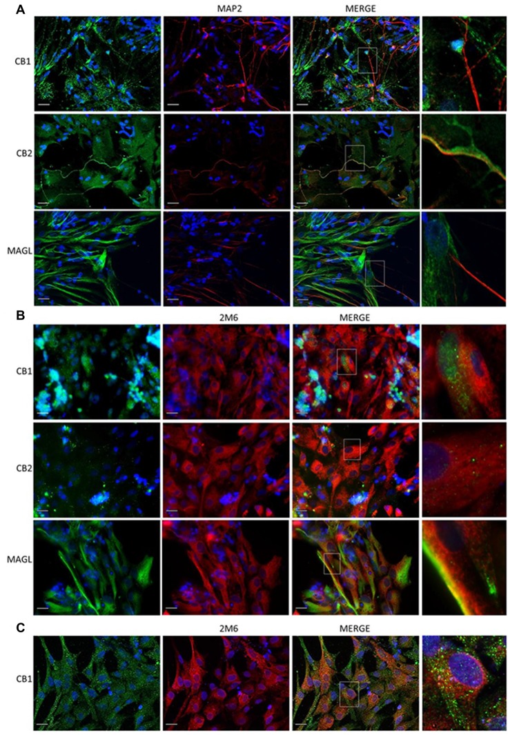
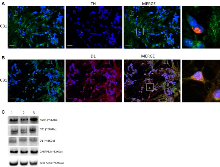
The avian retina has been used as a model to study signaling by different neuro- and gliotransmitters. It is unclear how dopaminergic and cannabinoid systems are related in the retina. Here we studied the expression of type 1 and 2 cannabinoid receptors (CB and CB), as well as monoacylglycerol lipase (MAGL), the enzyme that degrades 2-arachidonoylglycerol (2-AG), during retina development. Our data show that CB receptor is highly expressed from embryonic day 5 (E5) until post hatched day 7 (PE7), decreasing its levels throughout development. CB is densely found in the ganglion cell layer (GCL) and inner plexiform layer (IPL). CB receptor was also found from E5 until PE7 with a decrease in its contents from E9 afterwards. CB was mainly present in the lamination of the IPL at PE7. MAGL is expressed in all retinal layers, mainly in the IPL and OPL from E9 to PE7 retina. CB and CB were found both in neurons and glia cells, but MAGL was only expressed in Müller glia. Older retinas (PE7) show CB positive cells mainly in the INL and co-expression of CB and tyrosine hydroxylase (TH) are shown in a few cells when both systems are mature. CB co-localized with TH and was heavily associated to D receptor labeling in primary cell cultures. Finally, cyclic AMP (cAMP) was activated by the selective D agonist SKF38393, and inhibited when cultures were treated with WIN55, 212-2 (WIN) in a CB dependent manner. The results suggest a correlation between the endocannabinoid and dopaminergic systems (DSs) during the avian retina development. Activation of CB limits cAMP accumulation via D receptor activation and may influence embryological parameters during avian retina differentiation.
鸟类视网膜已被用作研究不同神经递质和神经胶质递质信号传导的模型。目前尚不清楚视网膜中多巴胺能系统和大麻素系统之间的关系。在此,我们研究了1型和2型大麻素受体(CB1和CB2)以及单酰甘油脂肪酶(MAGL,一种降解2-花生四烯酸甘油酯(2-AG)的酶)在视网膜发育过程中的表达。我们的数据表明,CB1受体从胚胎第5天(E5)到孵化后第7天(PE7)高度表达,在整个发育过程中其水平逐渐降低。CB2在神经节细胞层(GCL)和内网状层(IPL)中密集分布。CB1受体在E5到PE7期间也有表达,从E9之后其含量下降。CB2在PE7时主要存在于IPL的分层中。MAGL在所有视网膜层中均有表达,在E9到PE7的视网膜中主要存在于IPL和外网状层(OPL)。CB1和CB2在神经元和神经胶质细胞中均有发现,但MAGL仅在穆勒胶质细胞中表达。较成熟的视网膜(PE7)中,CB2阳性细胞主要在INL中,当两个系统成熟时,少数细胞中显示出CB2和酪氨酸羟化酶(TH)的共表达。在原代细胞培养中,CB2与TH共定位,并与D受体标记密切相关。最后,环磷酸腺苷(cAMP)被选择性D受体激动剂SKF38393激活,而当用WIN55,212-2(WIN)以CB依赖的方式处理培养物时,cAMP受到抑制。结果表明在鸟类视网膜发育过程中内源性大麻素系统和多巴胺能系统(DSs)之间存在相关性。CB1的激活通过D受体激活限制cAMP积累,并可能影响鸟类视网膜分化过程中的胚胎学参数。