• 文献检索
  • 文档翻译
  • 深度研究
  • 学术资讯
  • Suppr Zotero 插件Zotero 插件
  • 邀请有礼
  • 套餐&价格
  • 历史记录
应用&插件
Suppr Zotero 插件Zotero 插件浏览器插件Mac 客户端Windows 客户端微信小程序
定价
高级版会员购买积分包购买API积分包
服务
文献检索文档翻译深度研究API 文档MCP 服务
关于我们
关于 Suppr公司介绍联系我们用户协议隐私条款
关注我们

Suppr 超能文献

核心技术专利:CN118964589B侵权必究
粤ICP备2023148730 号-1Suppr @ 2026

文献检索

告别复杂PubMed语法,用中文像聊天一样搜索,搜遍4000万医学文献。AI智能推荐,让科研检索更轻松。

立即免费搜索

文件翻译

保留排版,准确专业,支持PDF/Word/PPT等文件格式,支持 12+语言互译。

免费翻译文档

深度研究

AI帮你快速写综述,25分钟生成高质量综述,智能提取关键信息,辅助科研写作。

立即免费体验

运动模型超声定位显微镜用于临床前和临床多参数肿瘤特征描述。

Motion model ultrasound localization microscopy for preclinical and clinical multiparametric tumor characterization.

机构信息

Institute for Experimental Molecular Imaging, University Clinic Aachen, RWTH Aachen University, CMBS, Forckenbeckstr. 55, 52074, Aachen, Germany.

Chair for Medical Engineering, Department of Electrical Engineering and Information Technology, Ruhr University Bochum, Universitätsstr. 150, 44780, Bochum, Germany.

出版信息

Nat Commun. 2018 Apr 18;9(1):1527. doi: 10.1038/s41467-018-03973-8.

DOI:10.1038/s41467-018-03973-8
PMID:29670096
原文链接:https://pmc.ncbi.nlm.nih.gov/articles/PMC5906644/
Abstract

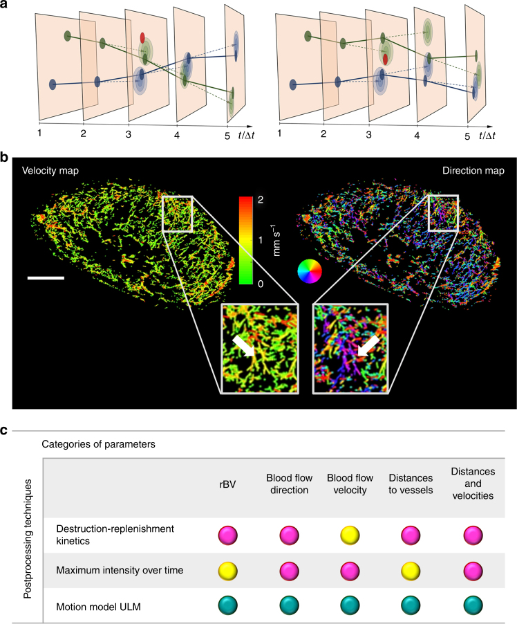
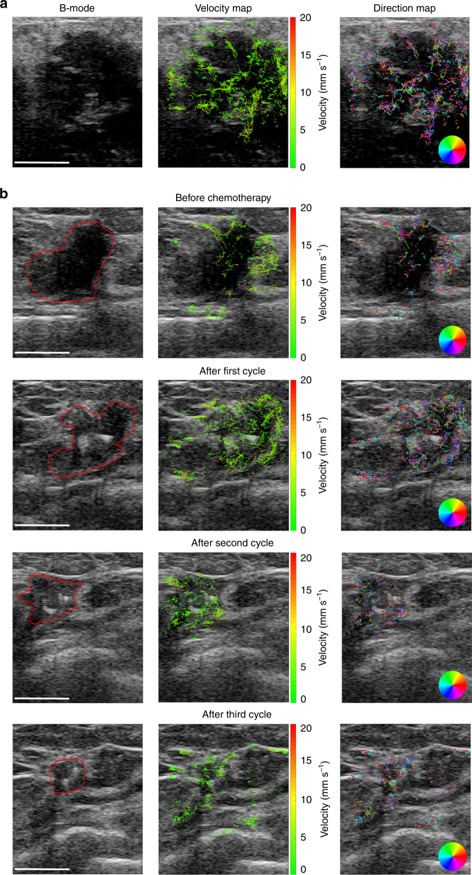
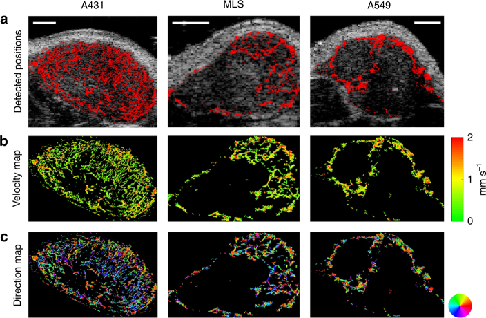
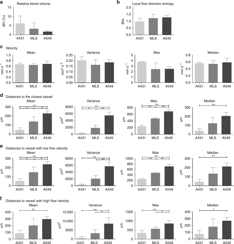
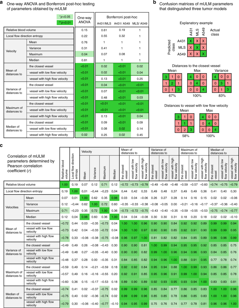
Super-resolution imaging methods promote tissue characterization beyond the spatial resolution limits of the devices and bridge the gap between histopathological analysis and non-invasive imaging. Here, we introduce motion model ultrasound localization microscopy (mULM) as an easily applicable and robust new tool to morphologically and functionally characterize fine vascular networks in tumors at super-resolution. In tumor-bearing mice and for the first time in patients, we demonstrate that within less than 1 min scan time mULM can be realized using conventional preclinical and clinical ultrasound devices. In this context, next to highly detailed images of tumor microvascularization and the reliable quantification of relative blood volume and perfusion, mULM provides multiple new functional and morphological parameters that discriminate tumors with different vascular phenotypes. Furthermore, our initial patient data indicate that mULM can be applied in a clinical ultrasound setting opening avenues for the multiparametric characterization of tumors and the assessment of therapy response.

摘要

超分辨率成像方法可促进组织特征分析超越设备的空间分辨率限制,并弥合组织病理学分析与非侵入性成像之间的差距。在这里,我们介绍运动模型超声定位显微镜 (mULM),这是一种易于应用且稳健的新工具,可在超分辨率下对肿瘤中的精细血管网络进行形态和功能特征分析。在荷瘤小鼠中,我们首次在患者中证明,使用传统的临床前和临床超声设备,不到 1 分钟的扫描时间即可实现 mULM。在这种情况下,除了肿瘤微血管化的高度详细图像以及相对血液体积和灌注的可靠定量之外,mULM 还提供了多个新的功能和形态参数,可以区分具有不同血管表型的肿瘤。此外,我们的初步患者数据表明,mULM 可应用于临床超声环境中,为肿瘤的多参数特征分析和治疗效果评估开辟了新途径。

https://cdn.ncbi.nlm.nih.gov/pmc/blobs/cca2/5906644/c078b0fd8672/41467_2018_3973_Fig6_HTML.jpg
https://cdn.ncbi.nlm.nih.gov/pmc/blobs/cca2/5906644/4eeb3cc56921/41467_2018_3973_Fig1_HTML.jpg
https://cdn.ncbi.nlm.nih.gov/pmc/blobs/cca2/5906644/771b03495e25/41467_2018_3973_Fig2_HTML.jpg
https://cdn.ncbi.nlm.nih.gov/pmc/blobs/cca2/5906644/455c6379cad1/41467_2018_3973_Fig3_HTML.jpg
https://cdn.ncbi.nlm.nih.gov/pmc/blobs/cca2/5906644/67520b87396b/41467_2018_3973_Fig4_HTML.jpg
https://cdn.ncbi.nlm.nih.gov/pmc/blobs/cca2/5906644/126d55ea9439/41467_2018_3973_Fig5_HTML.jpg
https://cdn.ncbi.nlm.nih.gov/pmc/blobs/cca2/5906644/c078b0fd8672/41467_2018_3973_Fig6_HTML.jpg

相似文献

1
Motion model ultrasound localization microscopy for preclinical and clinical multiparametric tumor characterization.运动模型超声定位显微镜用于临床前和临床多参数肿瘤特征描述。
Nat Commun. 2018 Apr 18;9(1):1527. doi: 10.1038/s41467-018-03973-8.
2
Ultrasound localization microscopy to image and assess microvasculature in a rat kidney.超声定位显微镜成像和评估大鼠肾脏的微血管。
Sci Rep. 2017 Oct 20;7(1):13662. doi: 10.1038/s41598-017-13676-7.
3
SUSHI: Sparsity-Based Ultrasound Super-Resolution Hemodynamic Imaging.基于稀疏的超声超分辨率血流成像。
IEEE Trans Ultrason Ferroelectr Freq Control. 2018 Dec;65(12):2365-2380. doi: 10.1109/TUFFC.2018.2873380. Epub 2018 Oct 2.
4
Detection and Tracking of Multiple Microbubbles in Ultrasound B-Mode Images.超声B模式图像中多个微泡的检测与跟踪
IEEE Trans Ultrason Ferroelectr Freq Control. 2016 Jan;63(1):72-82. doi: 10.1109/TUFFC.2015.2500266. Epub 2015 Nov 17.
5
Quantification of bound microbubbles in ultrasound molecular imaging.超声分子成像中结合微泡的定量分析。
IEEE Trans Ultrason Ferroelectr Freq Control. 2015 Jun;62(6):1190-200. doi: 10.1109/TUFFC.2015.006264.
6
Contrast-assisted destruction-replenishment ultrasound for the assessment of tumor microvasculature in a rat model.对比增强型破坏-补充超声用于评估大鼠模型中的肿瘤微血管。
Technol Cancer Res Treat. 2002 Dec;1(6):459-70. doi: 10.1177/153303460200100606.
7
In vivo acoustic super-resolution and super-resolved velocity mapping using microbubbles.利用微泡进行体内声超分辨率和超分辨率速度成像。
IEEE Trans Med Imaging. 2015 Feb;34(2):433-40. doi: 10.1109/TMI.2014.2359650. Epub 2014 Sep 23.
8
Ultrafast ultrasound localization microscopy for deep super-resolution vascular imaging.超快超声定位显微镜用于深层超分辨率血管成像。
Nature. 2015 Nov 26;527(7579):499-502. doi: 10.1038/nature16066.
9
Quantitative tissue perfusion imaging using nonlinear ultrasound localization microscopy.利用非线性超声定位显微镜进行定量组织灌注成像。
Sci Rep. 2022 Dec 19;12(1):21943. doi: 10.1038/s41598-022-24986-w.
10
Nanodroplet-Based Super-Resolution Ultrasound Localization Microscopy.基于纳米液滴的超分辨率超声定位显微镜
ACS Sens. 2023 Sep 22;8(9):3294-3306. doi: 10.1021/acssensors.3c00418. Epub 2023 Aug 22.

引用本文的文献

1
Current status and advances in ultrasound evaluation of neovascularization within carotid artery plaques: a systematic review.颈动脉斑块内新生血管形成的超声评估现状与进展:一项系统综述
Cardiovasc Ultrasound. 2025 Sep 1;23(1):19. doi: 10.1186/s12947-025-00356-0.
2
Comparison of the efficacy of FITC-conjugated dextran, PEG, and HA as intraoperative navigation materials in pancreatic cancer.异硫氰酸荧光素偶联葡聚糖、聚乙二醇和透明质酸作为胰腺癌术中导航材料的疗效比较。
RSC Adv. 2025 Aug 19;15(36):29300-29310. doi: 10.1039/d5ra02236c. eCollection 2025 Aug 18.
3
Ultrasound matrix imaging for 3D transcranial in vivo localization microscopy.

本文引用的文献

1
Automated Generation of Reliable Blood Velocity Parameter Maps from Contrast-Enhanced Ultrasound Data.从对比增强超声数据中自动生成可靠的血流速度参数图。
Contrast Media Mol Imaging. 2017 May 30;2017:2098324. doi: 10.1155/2017/2098324. eCollection 2017.
2
Ultrasound localization microscopy to image and assess microvasculature in a rat kidney.超声定位显微镜成像和评估大鼠肾脏的微血管。
Sci Rep. 2017 Oct 20;7(1):13662. doi: 10.1038/s41598-017-13676-7.
3
Subwavelength motion-correction for ultrafast ultrasound localization microscopy.
用于三维经颅体内定位显微镜检查的超声矩阵成像
Sci Adv. 2025 Aug;11(31):eadt9778. doi: 10.1126/sciadv.adt9778. Epub 2025 Jul 30.
4
Quantitative microvessel orientation biomarkers derived from contrast free ultrasound imaging for cancer diagnosis.源自无造影剂超声成像的定量微血管取向生物标志物用于癌症诊断。
Sci Rep. 2025 Jul 8;15(1):24500. doi: 10.1038/s41598-025-09745-x.
5
Advanced ultrasound methods to improve chronic kidney disease diagnosis.用于改善慢性肾脏病诊断的先进超声方法。
Npj Imaging. 2024 Jul 25;2(1):22. doi: 10.1038/s44303-024-00023-5.
6
Human lymph node microvascular imaging using a fast contrast-free super-resolution ultrasound technique.使用快速无造影剂超分辨率超声技术对人体淋巴结微血管进行成像。
Sci Rep. 2025 Jul 2;15(1):23061. doi: 10.1038/s41598-025-08483-4.
7
Analysing the Renal Vasculature Using Super-Resolution Ultrasound Imaging: Considerations for Clinical and Research Applications.使用超分辨率超声成像分析肾血管系统:临床和研究应用的注意事项
Diagnostics (Basel). 2025 Jun 14;15(12):1515. doi: 10.3390/diagnostics15121515.
8
Differentiation of benign and malignant breast lesions by ultrasound localization microscopy.超声定位显微镜对乳腺良恶性病变的鉴别诊断
Insights Imaging. 2025 Jun 18;16(1):128. doi: 10.1186/s13244-025-02013-6.
9
Progresses and clinical application of super-resolution ultrasound imaging: a narrative review.超分辨率超声成像的进展与临床应用:一篇叙述性综述
Ultrasound J. 2025 Jun 16;17(1):29. doi: 10.1186/s13089-025-00432-6.
10
Multiparametric super-resolution optoacoustic imaging with ICG-tagged microbubbles.使用吲哚菁绿标记微泡的多参数超分辨率光声成像
Mater Today Bio. 2025 May 12;32:101865. doi: 10.1016/j.mtbio.2025.101865. eCollection 2025 Jun.
用于超快超声定位显微镜的亚波长运动校正
Ultrasonics. 2017 May;77:17-21. doi: 10.1016/j.ultras.2017.01.008. Epub 2017 Jan 18.
4
3-D Ultrasound Localization Microscopy for Identifying Microvascular Morphology Features of Tumor Angiogenesis at a Resolution Beyond the Diffraction Limit of Conventional Ultrasound.用于在超越传统超声衍射极限的分辨率下识别肿瘤血管生成微血管形态特征的三维超声定位显微镜检查
Theranostics. 2017 Jan 1;7(1):196-204. doi: 10.7150/thno.16899. eCollection 2017.
5
Imaging in pancreatic disease.胰腺疾病的影像学诊断。
Nat Rev Gastroenterol Hepatol. 2017 Feb;14(2):97-109. doi: 10.1038/nrgastro.2016.144. Epub 2016 Nov 9.
6
Imaging biomarker roadmap for cancer studies.癌症研究的影像生物标志物路线图。
Nat Rev Clin Oncol. 2017 Mar;14(3):169-186. doi: 10.1038/nrclinonc.2016.162. Epub 2016 Oct 11.
7
Three-dimensional Contrast-enhanced Ultrasound in Response Assessment for Breast Cancer: A Comparison with Dynamic Contrast-enhanced Magnetic Resonance Imaging and Pathology.三维对比增强超声在乳腺癌疗效评估中的应用:与动态对比增强磁共振成像及病理学的比较
Sci Rep. 2016 Sep 22;6:33832. doi: 10.1038/srep33832.
8
Intra-Animal Comparison between Three-dimensional Molecularly Targeted US and Three-dimensional Dynamic Contrast-enhanced US for Early Antiangiogenic Treatment Assessment in Colon Cancer.结肠癌早期抗血管生成治疗评估中三维分子靶向超声与三维动态对比增强超声的动物体内比较
Radiology. 2017 Feb;282(2):443-452. doi: 10.1148/radiol.2016160032. Epub 2016 Aug 4.
9
Imalytics Preclinical: Interactive Analysis of Biomedical Volume Data.Imalytics临床前:生物医学体积数据的交互式分析
Theranostics. 2016 Jan 1;6(3):328-41. doi: 10.7150/thno.13624. eCollection 2016.
10
Ultrafast ultrasound localization microscopy for deep super-resolution vascular imaging.超快超声定位显微镜用于深层超分辨率血管成像。
Nature. 2015 Nov 26;527(7579):499-502. doi: 10.1038/nature16066.