• 文献检索
  • 文档翻译
  • 深度研究
  • 学术资讯
  • Suppr Zotero 插件Zotero 插件
  • 邀请有礼
  • 套餐&价格
  • 历史记录
应用&插件
Suppr Zotero 插件Zotero 插件浏览器插件Mac 客户端Windows 客户端微信小程序
定价
高级版会员购买积分包购买API积分包
服务
文献检索文档翻译深度研究API 文档MCP 服务
关于我们
关于 Suppr公司介绍联系我们用户协议隐私条款
关注我们

Suppr 超能文献

核心技术专利:CN118964589B侵权必究
粤ICP备2023148730 号-1Suppr @ 2026

文献检索

告别复杂PubMed语法,用中文像聊天一样搜索,搜遍4000万医学文献。AI智能推荐,让科研检索更轻松。

立即免费搜索

文件翻译

保留排版,准确专业,支持PDF/Word/PPT等文件格式,支持 12+语言互译。

免费翻译文档

深度研究

AI帮你快速写综述,25分钟生成高质量综述,智能提取关键信息,辅助科研写作。

立即免费体验

用于在超越传统超声衍射极限的分辨率下识别肿瘤血管生成微血管形态特征的三维超声定位显微镜检查

3-D Ultrasound Localization Microscopy for Identifying Microvascular Morphology Features of Tumor Angiogenesis at a Resolution Beyond the Diffraction Limit of Conventional Ultrasound.

作者信息

Lin Fanglue, Shelton Sarah E, Espíndola David, Rojas Juan D, Pinton Gianmarco, Dayton Paul A

机构信息

Joint Department of Biomedical Engineering, University of North Carolina at Chapel Hill and North Carolina State University, Chapel Hill, North Carolina, USA.

出版信息

Theranostics. 2017 Jan 1;7(1):196-204. doi: 10.7150/thno.16899. eCollection 2017.

DOI:10.7150/thno.16899
PMID:28042327
原文链接:https://pmc.ncbi.nlm.nih.gov/articles/PMC5196896/
Abstract

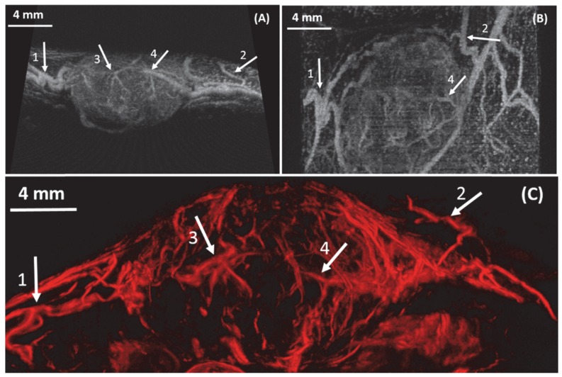
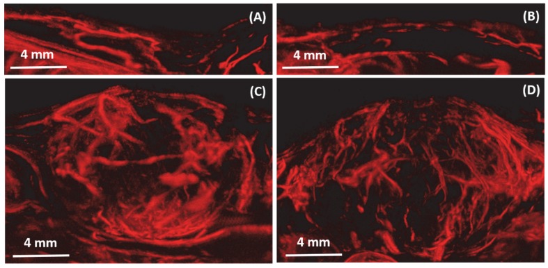
Angiogenesis has been known as a hallmark of solid tumor cancers for decades, yet ultrasound has been limited in its ability to detect the microvascular changes associated with malignancy. Here, we demonstrate the potential of 'ultrasound localization microscopy' applied volumetrically in combination with quantitative analysis of microvascular morphology, as an approach to overcome this limitation. This pilot study demonstrates our ability to image complex microvascular patterns associated with tumor angiogenesis in-vivo at a resolution of tens of microns - substantially better than the diffraction limit of traditional clinical ultrasound, yet using an 8 MHz clinical ultrasound probe. Furthermore, it is observed that data from healthy and tumor-bearing tissue exhibit significant differences in microvascular pattern and density. Results suggests that with continued development of these novel technologies, ultrasound has the potential to detect biomarkers of cancer based on the microvascular 'fingerprint' of malignant angiogenesis rather than through imaging of blood flow dynamics or the tumor mass itself.

摘要

几十年来,血管生成一直被视为实体肿瘤癌症的一个标志,然而超声在检测与恶性肿瘤相关的微血管变化方面能力有限。在此,我们展示了“超声定位显微镜”在体积上的应用潜力,并结合微血管形态的定量分析,以此作为克服这一局限性的方法。这项初步研究证明了我们能够以数十微米的分辨率在体内对与肿瘤血管生成相关的复杂微血管模式进行成像——这比传统临床超声的衍射极限要好得多,而且使用的是8兆赫的临床超声探头。此外,观察到来自健康组织和肿瘤组织的数据在微血管模式和密度上存在显著差异。结果表明,随着这些新技术的不断发展,超声有潜力基于恶性血管生成的微血管“指纹”而非通过血流动力学成像或肿瘤肿块本身来检测癌症生物标志物。

https://cdn.ncbi.nlm.nih.gov/pmc/blobs/7a2a/5196896/28619b3a037d/thnov07p0196g007.jpg
https://cdn.ncbi.nlm.nih.gov/pmc/blobs/7a2a/5196896/b5ecce827d5a/thnov07p0196g001.jpg
https://cdn.ncbi.nlm.nih.gov/pmc/blobs/7a2a/5196896/159cf06cd1d2/thnov07p0196g002.jpg
https://cdn.ncbi.nlm.nih.gov/pmc/blobs/7a2a/5196896/8f4ec4a75ddd/thnov07p0196g003.jpg
https://cdn.ncbi.nlm.nih.gov/pmc/blobs/7a2a/5196896/893dadf351df/thnov07p0196g004.jpg
https://cdn.ncbi.nlm.nih.gov/pmc/blobs/7a2a/5196896/d359b9fa6dab/thnov07p0196g005.jpg
https://cdn.ncbi.nlm.nih.gov/pmc/blobs/7a2a/5196896/5d4c7025b83d/thnov07p0196g006.jpg
https://cdn.ncbi.nlm.nih.gov/pmc/blobs/7a2a/5196896/28619b3a037d/thnov07p0196g007.jpg

相似文献

1
3-D Ultrasound Localization Microscopy for Identifying Microvascular Morphology Features of Tumor Angiogenesis at a Resolution Beyond the Diffraction Limit of Conventional Ultrasound.用于在超越传统超声衍射极限的分辨率下识别肿瘤血管生成微血管形态特征的三维超声定位显微镜检查
Theranostics. 2017 Jan 1;7(1):196-204. doi: 10.7150/thno.16899. eCollection 2017.
2
Evaluation of tumor microvasculature with 3D ultrasound localization microscopy based on 2D matrix array.基于二维矩阵阵列的三维超声定位显微镜对肿瘤微血管的评价。
Eur Radiol. 2024 Aug;34(8):5250-5259. doi: 10.1007/s00330-023-10039-x. Epub 2024 Jan 24.
3
The "Fingerprint" of Cancer Extends Beyond Solid Tumor Boundaries: Assessment With a Novel Ultrasound Imaging Approach.癌症的“指纹”超出实体瘤边界:采用新型超声成像方法进行评估
IEEE Trans Biomed Eng. 2016 May;63(5):1082-6. doi: 10.1109/TBME.2015.2479590. Epub 2015 Sep 18.
4
Early Assessment of Tumor Response to Radiation Therapy using High-Resolution Quantitative Microvascular Ultrasound Imaging.使用高分辨率定量微血管超声成像技术早期评估肿瘤对放射治疗的反应。
Theranostics. 2018 Jan 1;8(1):156-168. doi: 10.7150/thno.19703. eCollection 2018.
5
The implementation of acoustic angiography for microvascular and angiogenesis imaging.用于微血管和血管生成成像的声学血管造影术的实施。
Annu Int Conf IEEE Eng Med Biol Soc. 2014;2014:4283-5. doi: 10.1109/EMBC.2014.6944571.
6
Optimizing Sensitivity of Ultrasound Contrast-Enhanced Super-Resolution Imaging by Tailoring Size Distribution of Microbubble Contrast Agent.通过调整微泡造影剂的尺寸分布优化超声造影增强超分辨率成像的灵敏度
Ultrasound Med Biol. 2017 Oct;43(10):2488-2493. doi: 10.1016/j.ultrasmedbio.2017.05.014. Epub 2017 Jun 28.
7
Superharmonic Ultrasound for Motion-Independent Localization Microscopy: Applications to Microvascular Imaging From Low to High Flow Rates.用于运动独立定位显微镜的超声谐波:从低流速到高流速的微血管成像应用。
IEEE Trans Ultrason Ferroelectr Freq Control. 2020 May;67(5):957-967. doi: 10.1109/TUFFC.2020.2965767. Epub 2020 Jan 10.
8
Mapping microvasculature with acoustic angiography yields quantifiable differences between healthy and tumor-bearing tissue volumes in a rodent model.声学血管造影技术可绘制微血管图谱,在啮齿动物模型中可量化健康组织与肿瘤组织体积的差异。
Radiology. 2012 Sep;264(3):733-40. doi: 10.1148/radiol.12112000. Epub 2012 Jul 6.
9
3-D In Vitro Acoustic Super-Resolution and Super-Resolved Velocity Mapping Using Microbubbles.基于微泡的三维体外声超分辨率与超分辨率速度成像。
IEEE Trans Ultrason Ferroelectr Freq Control. 2017 Oct;64(10):1478-1486. doi: 10.1109/TUFFC.2017.2731664. Epub 2017 Jul 31.
10
On the Relationship between Dynamic Contrast-Enhanced Ultrasound Parameters and the Underlying Vascular Architecture Extracted from Acoustic Angiography.关于动态对比增强超声参数与从声学血管造影提取的潜在血管结构之间的关系
Ultrasound Med Biol. 2019 Feb;45(2):539-548. doi: 10.1016/j.ultrasmedbio.2018.08.018. Epub 2018 Nov 30.

引用本文的文献

1
Implicit neural representation for scalable 3D reconstruction from sparse ultrasound images.用于从稀疏超声图像进行可扩展三维重建的隐式神经表示
NPJ Acoust. 2025;1(1):14. doi: 10.1038/s44384-025-00018-5. Epub 2025 Aug 8.
2
Ultrasound matrix imaging for 3D transcranial in vivo localization microscopy.用于三维经颅体内定位显微镜检查的超声矩阵成像
Sci Adv. 2025 Aug;11(31):eadt9778. doi: 10.1126/sciadv.adt9778. Epub 2025 Jul 30.
3
Differentiation of benign and malignant breast lesions by ultrasound localization microscopy.超声定位显微镜对乳腺良恶性病变的鉴别诊断

本文引用的文献

1
Assessment of Molecular Acoustic Angiography for Combined Microvascular and Molecular Imaging in Preclinical Tumor Models.临床前肿瘤模型中用于微血管和分子联合成像的分子声学血管造影评估
Mol Imaging Biol. 2017 Apr;19(2):194-202. doi: 10.1007/s11307-016-0991-4.
2
Molecular Acoustic Angiography: A New Technique for High-resolution Superharmonic Ultrasound Molecular Imaging.分子声学血管造影术:一种用于高分辨率超谐波超声分子成像的新技术。
Ultrasound Med Biol. 2016 Mar;42(3):769-81. doi: 10.1016/j.ultrasmedbio.2015.10.015. Epub 2015 Dec 8.
3
Ultrafast ultrasound localization microscopy for deep super-resolution vascular imaging.
Insights Imaging. 2025 Jun 18;16(1):128. doi: 10.1186/s13244-025-02013-6.
4
Online 4D Ultrasound-Guided Robotic Tracking Enables 3D Ultrasound Localisation Microscopy with Large Tissue Displacements.在线4D超声引导机器人跟踪实现了具有大组织位移的3D超声定位显微镜检查。
IEEE Trans Med Imaging. 2025 May 19;PP:1. doi: 10.1109/TMI.2025.3571392.
5
Background Noise Removal in Non-Contrast- Enhanced Ultrasound Microvasculature Imaging Using Combined Collaborative, Morphological, and Vesselness Filtering.基于协同、形态学和血管性滤波相结合的非增强超声微血管成像背景噪声去除
IEEE Access. 2024;12:116150-116161. doi: 10.1109/access.2024.3446531. Epub 2024 Aug 20.
6
Achieving single cell acoustic localisation with deactivation super resolution.通过失活超分辨率实现单细胞声学定位。
NPJ Acoust. 2025;1(1):5. doi: 10.1038/s44384-025-00008-7. Epub 2025 Apr 24.
7
Optimizingdata acquisition for robust clinical microvascular imaging using ultrasound localization microscopy.利用超声定位显微镜优化数据采集以实现稳健的临床微血管成像。
Phys Med Biol. 2025 Mar 27;70(7). doi: 10.1088/1361-6560/adc0de.
8
Advancements in Three-Dimensional Super-Resolution Ultrasound Imaging: A Narrative Review.三维超分辨率超声成像的进展:一项叙述性综述。
J Ultrasound Med. 2025 Jul;44(7):1157-1174. doi: 10.1002/jum.16682. Epub 2025 Mar 12.
9
Ultrasound Super-Resolution Imaging of Neonatal Cerebral Vascular Reorganization.新生儿脑血管重组的超声超分辨率成像
Adv Sci (Weinh). 2025 Mar;12(12):e2415235. doi: 10.1002/advs.202415235. Epub 2025 Feb 3.
10
Ultrasound localization microscopy in the diagnosis of breast tumors and prediction of relevant histologic biomarkers associated with prognosis in humans: the protocol for a prospective, multicenter study.超声定位显微镜在人类乳腺肿瘤诊断及与预后相关的组织学生物标志物预测中的应用:一项前瞻性多中心研究方案
BMC Med Imaging. 2025 Jan 8;25(1):13. doi: 10.1186/s12880-024-01535-7.
超快超声定位显微镜用于深层超分辨率血管成像。
Nature. 2015 Nov 26;527(7579):499-502. doi: 10.1038/nature16066.
4
The "Fingerprint" of Cancer Extends Beyond Solid Tumor Boundaries: Assessment With a Novel Ultrasound Imaging Approach.癌症的“指纹”超出实体瘤边界:采用新型超声成像方法进行评估
IEEE Trans Biomed Eng. 2016 May;63(5):1082-6. doi: 10.1109/TBME.2015.2479590. Epub 2015 Sep 18.
5
Spatiotemporal Clutter Filtering of Ultrafast Ultrasound Data Highly Increases Doppler and fUltrasound Sensitivity.时空杂波滤波极大提高了超快速超声数据的多普勒和 f 超声灵敏度。
IEEE Trans Med Imaging. 2015 Nov;34(11):2271-85. doi: 10.1109/TMI.2015.2428634. Epub 2015 Apr 30.
6
Quantification of Microvascular Tortuosity during Tumor Evolution Using Acoustic Angiography.使用声学血管造影术对肿瘤进展过程中的微血管迂曲度进行量化分析。
Ultrasound Med Biol. 2015 Jul;41(7):1896-904. doi: 10.1016/j.ultrasmedbio.2015.02.012. Epub 2015 Apr 7.
7
In vivo acoustic super-resolution and super-resolved velocity mapping using microbubbles.利用微泡进行体内声超分辨率和超分辨率速度成像。
IEEE Trans Med Imaging. 2015 Feb;34(2):433-40. doi: 10.1109/TMI.2014.2359650. Epub 2014 Sep 23.
8
A super-resolution ultrasound method for brain vascular mapping.一种用于脑血管测绘的超高分辨率超声方法。
Med Phys. 2013 Nov;40(11):110701. doi: 10.1118/1.4823762.
9
Ultrasound contrast imaging: influence of scatterer motion in multi-pulse techniques.超声对比成像:多脉冲技术中散射体运动的影响。
IEEE Trans Ultrason Ferroelectr Freq Control. 2013 Oct;60(10):2065-78. doi: 10.1109/TUFFC.2013.2797.
10
Acoustic angiography: a new imaging modality for assessing microvasculature architecture.声学血管造影术:一种评估微血管结构的新型成像方式。
Int J Biomed Imaging. 2013;2013:936593. doi: 10.1155/2013/936593. Epub 2013 Jul 17.