Department of Pathology and Laboratory Medicine, David Geffen School of Medicine at UCLA, Los Angeles, California, United States of America.
PLoS One. 2018 Apr 19;13(4):e0195958. doi: 10.1371/journal.pone.0195958. eCollection 2018.
Immune checkpoint regulators, cytotoxic T lymphocyte antigen 4 (CTLA-4) and the programmed cell death protein-1/programmed death-ligand 1 (PD-1/PD-L1) have emerged as promising new targets for cancer therapeutics. While tumor expression of PD-L1 has been shown to have objective responses to anti-PD-L1 immunotherapies, the clinical implications of CTLA-4 expression in tumor cells or immune cells in the tumor microenvironment is still controversial. We investigated the expression of CTLA-4 and PD-L1 in human breast tumors and provided a scoring system for the systematic evaluation of CTLA-4 staining.
Immunohistochemical staining for PD-L1 and CTLA-4 expression was performed on a tissue microarray of 102 cores, which included normal and neoplastic breast tissues. Neoplastic cores were divided into four groups: Ductal carcinoma in situ (DCIS), invasive ductal carcinoma (IDC), invasive lobular carcinoma (ILC) and invasive tubular carcinoma (ITC). PD-L1 and CTLA-4 expressions were scored based on a system which accounted for the percentage and intensity of positivity and results provided in conjunction with available clinical and demographic data.
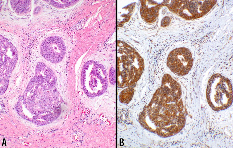
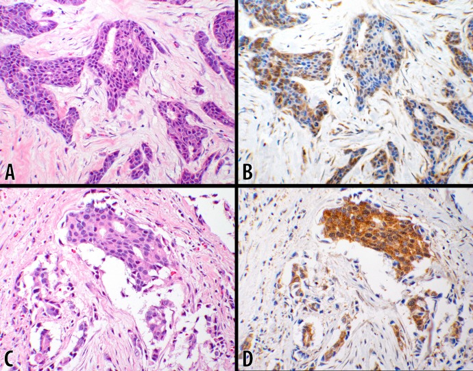
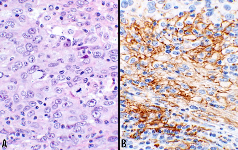
Overall, CTLA-4 was over-expressed in 49 of 93 (52.7%) breast tumors. Subcategorically, CTLA-4 was positive in 3 of 8 (37.5%) ductal carcinoma in situ, 40 of 73 (55%) of invasive ductal carcinomas, 4 of 10 (40%) of invasive lobular carcinomas and 2 of 2 (100%) of invasive tubular carcinomas. All 6 normal breast tissues were interpreted as negative for CTLA-4 staining. Only 4.1% of the invasive ductal carcinomas were positive for PD-L1 reactivity and the remaining carcinomas stained negative.
This study shows a significant overexpression of CTLA-4 in >50% of breast carcinomas with no such overexpression of CTLA-4 in benign breast tissues. PDL-1 staining is seen in only a small number of invasive ductal carcinomas (4.1%). These findings suggest the need for further investigation of anti-CTLA-4 and anti-PD-L1 immunotherapies and their efficacy in the treatment of breast carcinomas with overexpression of these immune modulators. In addition, the proposed scoring system will facilitate a more systematic correlation between tumor reactivity and clinical outcome which can be applied to all intracytoplasmic tumor markers.
免疫检查点调节剂、细胞毒性 T 淋巴细胞相关抗原 4(CTLA-4)和程序性细胞死亡蛋白 1/程序性死亡配体 1(PD-1/PD-L1)已成为癌症治疗的有前途的新靶点。虽然肿瘤 PD-L1 的表达与抗 PD-L1 免疫疗法的客观反应有关,但肿瘤细胞中 CTLA-4 的表达或肿瘤微环境中免疫细胞的临床意义仍存在争议。我们研究了 CTLA-4 和 PD-L1 在人乳腺癌中的表达,并提供了一种用于系统评估 CTLA-4 染色的评分系统。
对包括正常和肿瘤性乳腺组织的 102 个组织微阵列进行 PD-L1 和 CTLA-4 表达的免疫组织化学染色。肿瘤性核心分为四组:导管原位癌(DCIS)、浸润性导管癌(IDC)、浸润性小叶癌(ILC)和浸润性管状癌(ITC)。根据一种考虑阳性百分比和强度的系统对 PD-L1 和 CTLA-4 的表达进行评分,并结合可用的临床和人口统计学数据提供结果。
总体而言,93 例乳腺癌中有 49 例(52.7%)过度表达 CTLA-4。亚组分析显示,3 例(37.5%)导管原位癌、40 例(55%)浸润性导管癌、4 例(40%)浸润性小叶癌和 2 例(100%)浸润性管状癌中 CTLA-4 阳性。所有 6 例正常乳腺组织均被解读为 CTLA-4 染色阴性。只有 4.1%的浸润性导管癌对 PD-L1 反应阳性,其余的癌均为阴性。
本研究表明,超过 50%的乳腺癌中 CTLA-4 过度表达,而良性乳腺组织中无 CTLA-4 过度表达。只有少数浸润性导管癌(4.1%)出现 PD-L1 染色。这些发现表明需要进一步研究抗 CTLA-4 和抗 PD-L1 免疫疗法及其在这些免疫调节剂过度表达的乳腺癌中的疗效。此外,所提出的评分系统将有助于更系统地将肿瘤反应与临床结果相关联,可应用于所有细胞内肿瘤标志物。