Jin Xin, Zhang Man, Zhu Xue-Min, Fan Yan-Ru, Du Chen-Guang, Bao Hua-Er, Xu Siri-Guleng, Tian Qiao-Zhen, Wang Yun-He, Yang Yin-Feng
Veterinary Medicine College of Inner Mongolia Agricultural University, Hohhot, 010018, People's Republic of China.
Key Laboratory of Clinical Diagnosis and Treatment Technology in Animal Disease, Ministry of Agriculture, Hohhot, 010018, People's Republic of China.
BMC Vet Res. 2018 Apr 19;14(1):134. doi: 10.1186/s12917-018-1445-9.
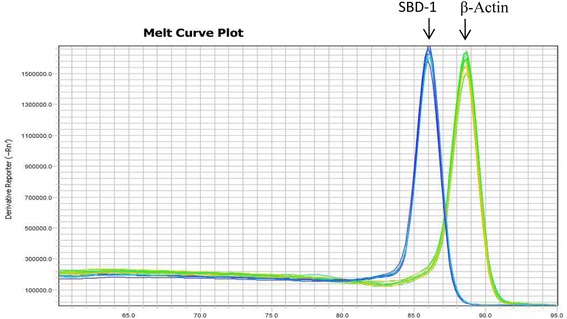
The ovine rumen is involved in host defense responses and acts as the immune interface with the environment. The ruminal mucosal epithelium plays an important role in innate immunity and secretes antimicrobial innate immune molecules that have bactericidal activity against a variety of pathogens. Defensins are cationic peptides that are produced by the mucosal epithelia and have broad-spectrum antimicrobial activity. Sheep β-defensin-1 (SBD-1) is one of the most important antibacterial peptides in the rumen. The expression of SBD-1 is regulated by the probiotic, Saccharomyces cerevisiae (S.c); however, the regulatory mechanism has not yet been elucidated. In the current study, the effects of S.c on the expression and secretion of SBD-1 in ovine ruminal epithelial cells were investigated using quantitative real-time PCR (qPCR) and enzyme-linked immunosorbent assay (ELISA). In addition, specific inhibitors were used to block the nuclear factor kappa-light-chain enhancer of activated B cells (NF-κB), p38, JNK, and ERK1/2 signalling pathways separately or simultaneously, to determine the regulatory mechanism(s) governing S.c-induced SBD-1 upregulation.
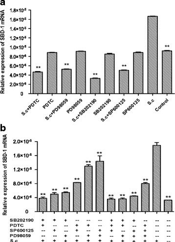
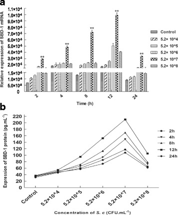
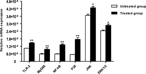
Incubation with S.c induced release of SBD-1 by ovine ruminal epithelial cells, with SBD-1 expression peaking after 12 h of incubation. The highest SBD-1 expression levels were achieved after treatment with 5.2 × 10 CFU∙mL S.c. Treatment with S.c resulted in significantly increased NF-κB, p38, JNK, ERK1/2, TLR2, and MyD88 mRNA expression. Whereas inhibition of mitogen-activated protein kinases (MAPKs) and NF-κB gene expression led to a decrease in SBD-1 expression.
S.c was induced SBD-1 expression and the S.c-induced up-regulation of SBD-1 expression may be related to TLR2 and MyD88 in ovine ruminal epithelial cells. This is likely simultaneously regulated by the MAPKs and NF-κB pathways with the p38 axis of the MAPKs pathway acting as the primary regulator. Thus, the pathways regulating S.c-induced SBD-1 expression may be related to TLR2-MyD88-NF-κB/MAPKs, with the TLR2-MyD88-p38 component of the TLR2-MyD88-MAPKs signalling acting as the main pathway.
绵羊瘤胃参与宿主防御反应,是与外界环境的免疫界面。瘤胃黏膜上皮在固有免疫中起重要作用,可分泌对多种病原体具有杀菌活性的抗菌固有免疫分子。防御素是由黏膜上皮产生的阳离子肽,具有广谱抗菌活性。绵羊β-防御素-1(SBD-1)是瘤胃中最重要的抗菌肽之一。SBD-1的表达受益生菌酿酒酵母(S.c)调控,但其调控机制尚未阐明。在本研究中,采用定量实时PCR(qPCR)和酶联免疫吸附测定(ELISA)研究了S.c对绵羊瘤胃上皮细胞中SBD-1表达和分泌的影响。此外,使用特异性抑制剂分别或同时阻断活化B细胞的核因子κB轻链增强子(NF-κB)、p38、JNK和ERK1/2信号通路,以确定调控S.c诱导SBD-1上调的机制。
与S.c共孵育可诱导绵羊瘤胃上皮细胞释放SBD-1,孵育12小时后SBD-1表达达到峰值。用5.2×10 CFU∙mL S.c处理后,SBD-1表达水平最高。用S.c处理导致NF-κB、p38、JNK、ERK1/2、TLR2和MyD88 mRNA表达显著增加。而抑制丝裂原活化蛋白激酶(MAPKs)和NF-κB基因表达导致SBD-1表达下降。
S.c诱导SBD-1表达,S.c诱导的SBD-1表达上调可能与绵羊瘤胃上皮细胞中的TLR2和MyD88有关。这可能同时受MAPKs和NF-κB通路调控,其中MAPKs通路的p38轴起主要调节作用。因此,调控S.c诱导SBD-1表达的通路可能与TLR2-MyD88-NF-κB/MAPKs有关,TLR2-MyD88-MAPKs信号的TLR2-MyD88-p38成分起主要通路作用。