Department of Cardiology, The Second Affiliated Hospital of Fujian Medical University, Quanzhou, 362000, Fujian, People's Republic of China.
The Key Laboratory of Cardiovascular Remodeling and Function Research, Chinese Ministry of Education and Chinese Ministry of Health, The State and Shandong Province Joint Key Laboratory of Translational Cardiovascular Medicine, Department of Cardiology, Qilu Hospital of Shandong University, No. 107, Wen Hua Xi Road, Jinan, 250012, Shandong, China.
J Transl Med. 2018 Apr 19;16(1):106. doi: 10.1186/s12967-018-1479-6.
Intermittent hypoxia (IH), a typical character of obstructive sleep apnea (OSA), is related to atherogenesis. However, the role of IH on atherosclerosis (AS) progression and the mechanisms involved remains poorly understood.
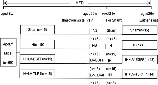
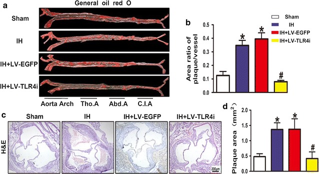
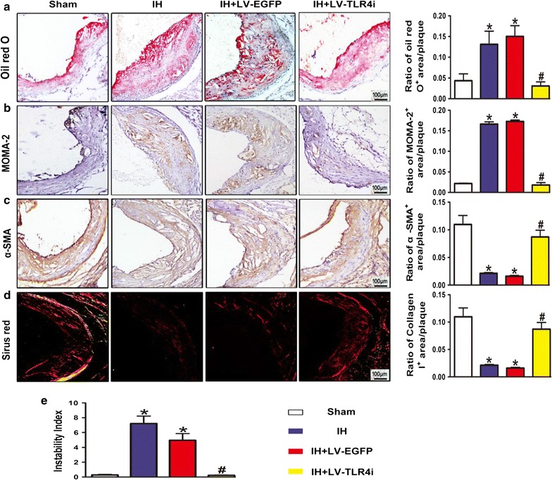
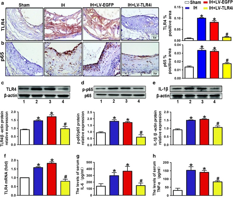
In the present study, high-fat fed ApoE mice were treated with recombinant shRNA-TLR4 lentivirus and exposed to IH. Atherosclerotic lesions on the en face aorta and cross-sections of aortic root were examined by Oil-Red O staining. The content of lipids and collagen of aortic root plaques were detected by Oil-Red O staining and Sirius red staining, respectively. The TLR4, NF-κB p65, α-SMA and MOMA-2 expression in aorta and IL-6 and TNF-α expression in the mice serum were also detected.
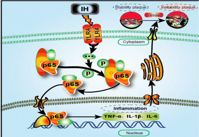
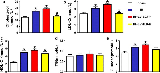
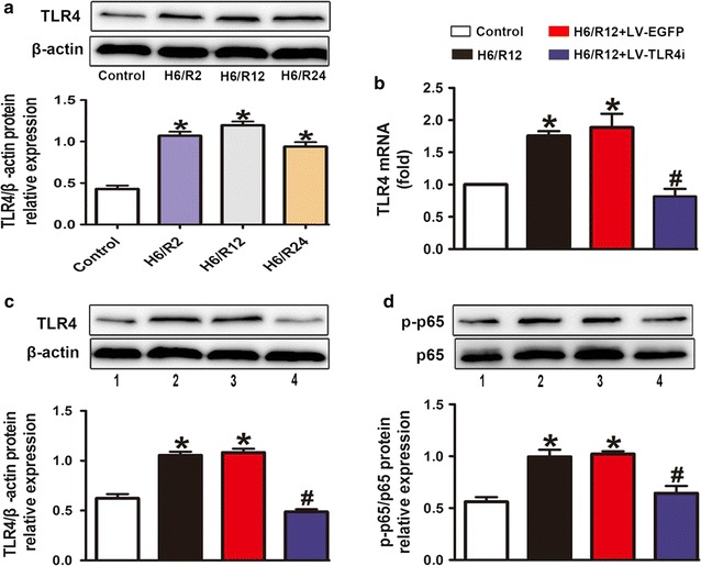
Compared with the Sham group, the IH treated group further increased atherosclerotic plaque loads and plaque vulnerability in the aortic sinus. Along with increased TLR4 expression, enhanced NF-κB activation, inflammatory activity and aggravated dyslipidemia were observed in the IH treated group. TLR4 interference partly inhibited IH-mediated AS progression with decreased inflammation and improved cholesterol levels. Similarly, in endothelial cells, hypoxia/reoxygenation exposure has been shown to promote TLR4 expression and activation of proinflammatory TLR4/NF-κB signaling, while TLR4 interference inhibited these effects.
We found that the IH accelerated growth and vulnerability of atherosclerotic plaque, which probably acted by triggering the activation of proinflammatory TLR4/NF-κB signaling. These findings may suggest that IH is a risk factor for vulnerable plaque and provide a new insight into the treatment of OSA-induced AS progression.
间歇性低氧(IH)是阻塞性睡眠呼吸暂停(OSA)的典型特征,与动脉粥样硬化(AS)的形成有关。然而,IH 对动脉粥样硬化进展的作用及其相关机制仍知之甚少。
在本研究中,高脂喂养的载脂蛋白 E(ApoE)基因敲除小鼠接受重组 shRNA-TLR4 慢病毒治疗,并暴露于 IH 环境中。采用油红 O 染色法检测主动脉正面和主动脉根部横切片的动脉粥样硬化病变。油红 O 染色和天狼猩红染色分别检测主动脉根部斑块的脂质和胶原含量。还检测了主动脉 TLR4、NF-κB p65、α-SMA 和 MOMA-2 的表达以及小鼠血清中 IL-6 和 TNF-α 的表达。
与 Sham 组相比,IH 处理组进一步增加了主动脉窦的动脉粥样硬化斑块负荷和斑块易损性。IH 处理组 TLR4 表达增加,NF-κB 激活增强,炎症活性增加,血脂异常加重。TLR4 干扰部分抑制了 IH 介导的 AS 进展,炎症减少,胆固醇水平改善。同样,在血管内皮细胞中,缺氧/复氧暴露已被证明可促进 TLR4 表达和促炎 TLR4/NF-κB 信号通路的激活,而 TLR4 干扰抑制了这些作用。
我们发现 IH 加速了动脉粥样硬化斑块的生长和易损性,这可能是通过触发促炎 TLR4/NF-κB 信号通路的激活来实现的。这些发现可能表明 IH 是易损斑块的一个危险因素,并为治疗 OSA 引起的 AS 进展提供了新的思路。