• 文献检索
  • 文档翻译
  • 深度研究
  • 学术资讯
  • Suppr Zotero 插件Zotero 插件
  • 邀请有礼
  • 套餐&价格
  • 历史记录
应用&插件
Suppr Zotero 插件Zotero 插件浏览器插件Mac 客户端Windows 客户端微信小程序
定价
高级版会员购买积分包购买API积分包
服务
文献检索文档翻译深度研究API 文档MCP 服务
关于我们
关于 Suppr公司介绍联系我们用户协议隐私条款
关注我们

Suppr 超能文献

核心技术专利:CN118964589B侵权必究
粤ICP备2023148730 号-1Suppr @ 2026

文献检索

告别复杂PubMed语法,用中文像聊天一样搜索,搜遍4000万医学文献。AI智能推荐,让科研检索更轻松。

立即免费搜索

文件翻译

保留排版,准确专业,支持PDF/Word/PPT等文件格式,支持 12+语言互译。

免费翻译文档

深度研究

AI帮你快速写综述,25分钟生成高质量综述,智能提取关键信息,辅助科研写作。

立即免费体验

坐骨肛门窝孤立性纤维瘤——一种结合放射学与病理学相关性的多学科管理方法

Solitary fibrous tumor of the ischioanal fossa-a multidisciplinary approach to management with radiologic-pathologic correlation.

作者信息

Bhat A, Layfield L J, Tewari S O, Gaballah A H, Davis R, Wu Z

机构信息

Department of Diagnostic Radiology, Interventional Radiology Section, University of Missouri, 1 Hospital Drive, Columbia, MO 65201, USA.

Department of Pathology and Anatomical Sciences, University of Missouri, 1 Hospital Drive, Columbia, MO 65201, USA.

出版信息

Radiol Case Rep. 2018 Mar 2;13(2):468-474. doi: 10.1016/j.radcr.2018.01.030. eCollection 2018 Apr.

DOI:10.1016/j.radcr.2018.01.030
PMID:29682137
原文链接:https://pmc.ncbi.nlm.nih.gov/articles/PMC5906773/
Abstract

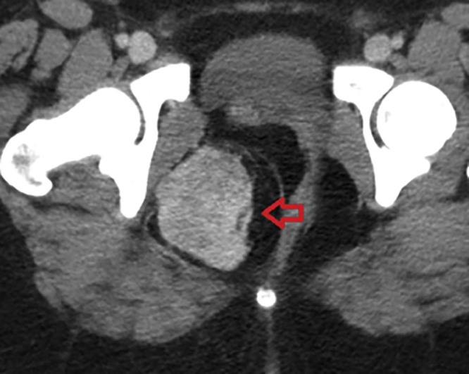
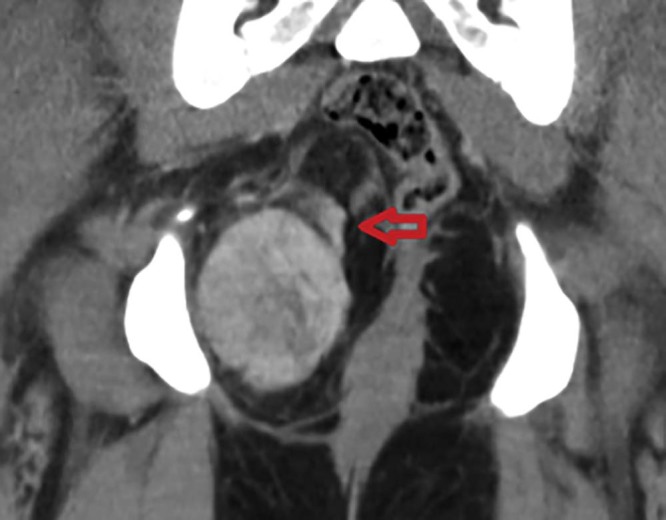
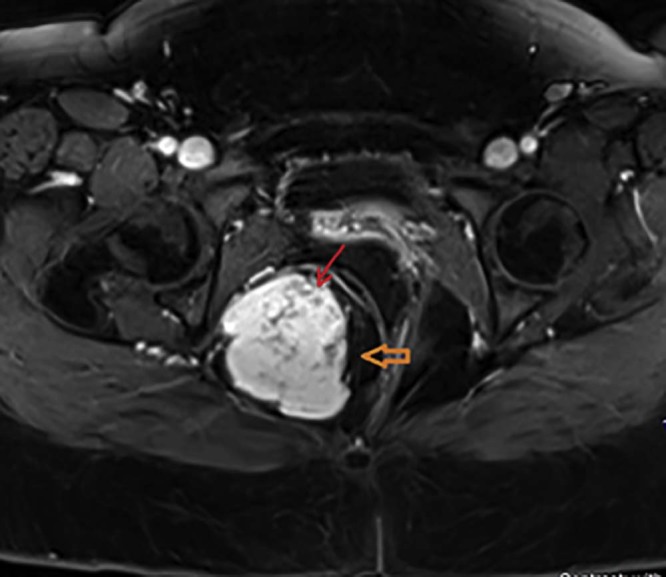
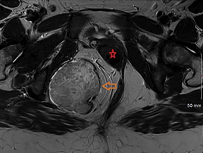
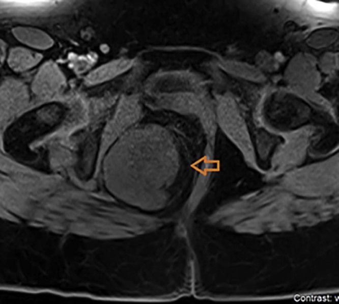
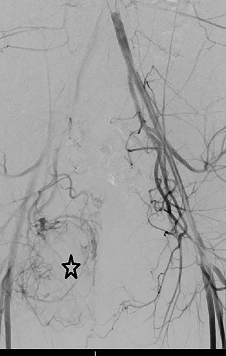
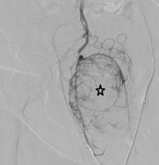
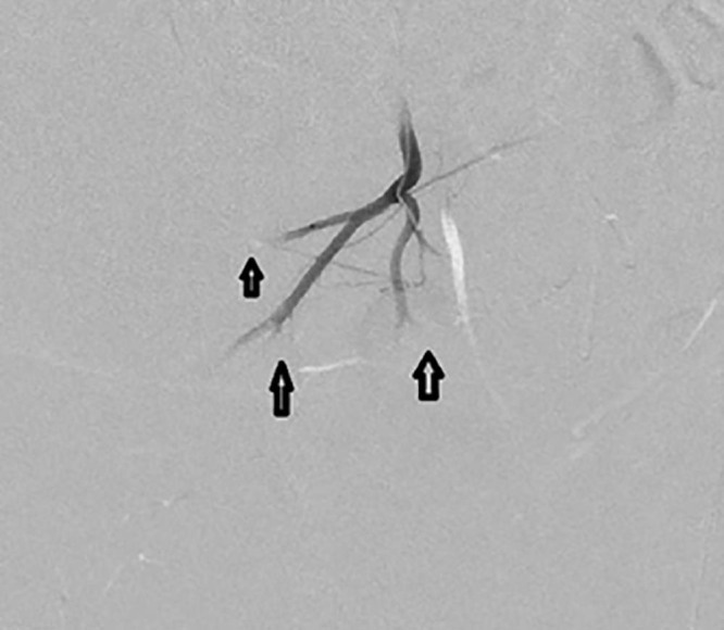
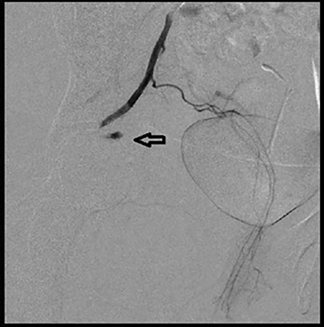
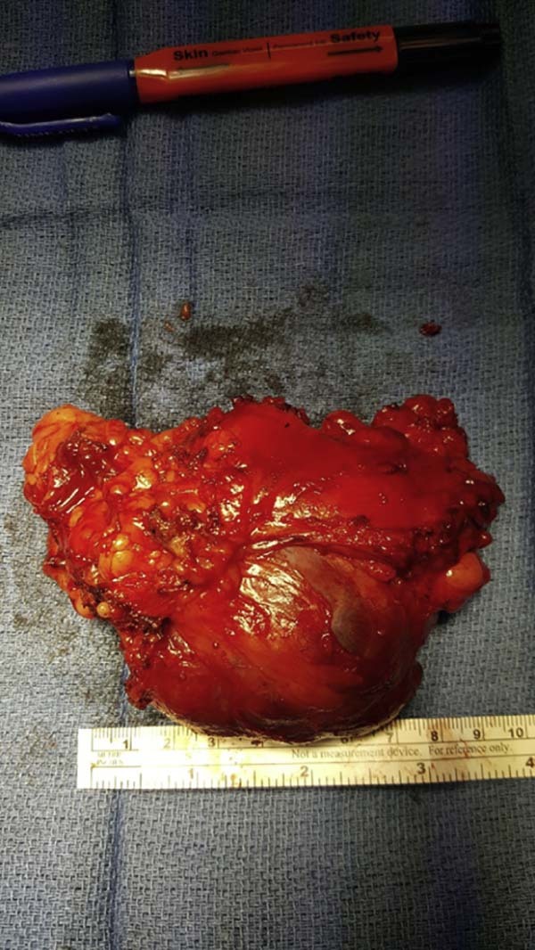
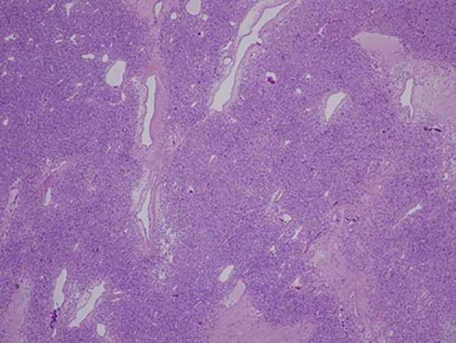
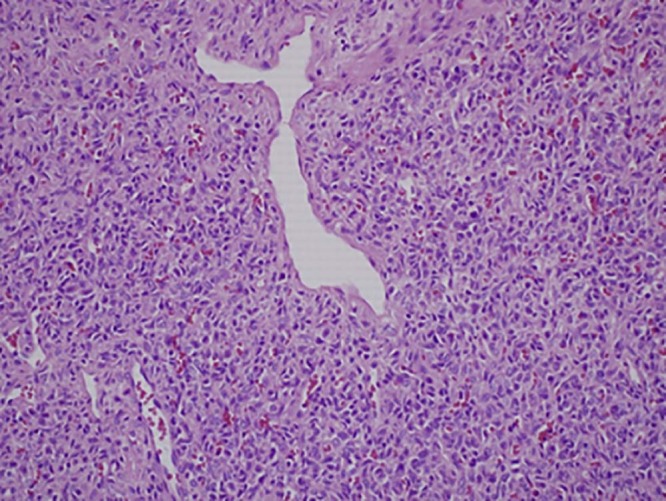
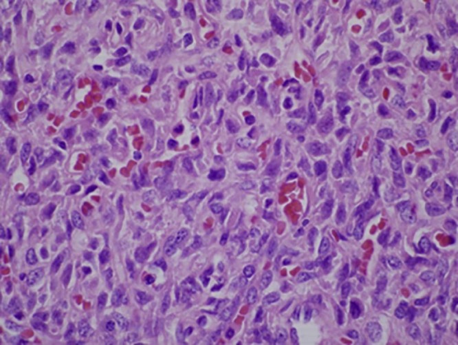
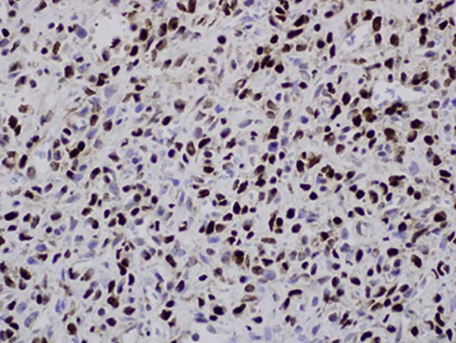
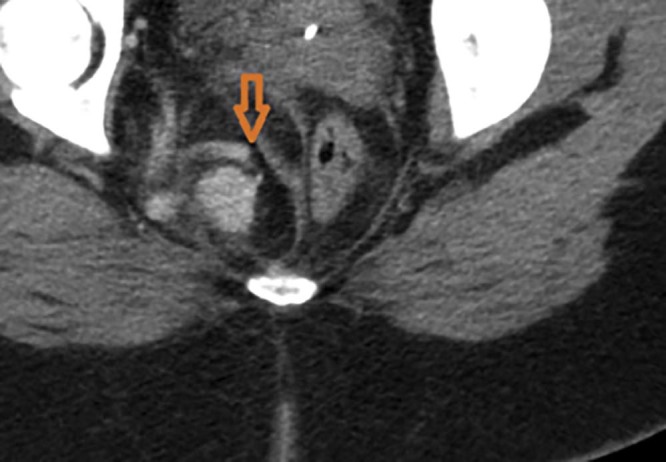
Solitary fibrous tumors are primary mesenchymal tumors, which may occur in any part of the body. Overall, these tumors are considered to have intermediate malignant potential with 5- and 10-year metastasis-free and overall disease-specific survival rates of 74% and 55%, and 89% and 73%, respectively (Demicco et al, 2012). Herein we present an unusual case of solitary fibrous tumors involving the ischioanal fossa in a 19-year-old woman with radiologic-pathologic correlation. This case was complicated by extensive tumor vascularity and was thus managed with preoperative embolization followed by en bloc surgical resection.

摘要

孤立性纤维瘤是原发性间叶组织肿瘤,可发生于身体的任何部位。总体而言,这些肿瘤被认为具有中等恶性潜能,5年和10年无转移生存率及疾病特异性总生存率分别为74%和55%,以及89%和73%(德米科等人,2012年)。在此,我们报告一例罕见的孤立性纤维瘤病例,该病例发生于一名19岁女性的坐骨肛门窝,并进行了放射学与病理学相关性分析。该病例因肿瘤血管丰富而复杂化,因此先进行术前栓塞,然后行整块手术切除。

https://cdn.ncbi.nlm.nih.gov/pmc/blobs/ba8d/5906773/9d87cefb070d/radcr450-fig-0015.jpg
https://cdn.ncbi.nlm.nih.gov/pmc/blobs/ba8d/5906773/d6bca0cd2e48/radcr450-fig-0001.jpg
https://cdn.ncbi.nlm.nih.gov/pmc/blobs/ba8d/5906773/d1ef5df0f5b2/radcr450-fig-0002.jpg
https://cdn.ncbi.nlm.nih.gov/pmc/blobs/ba8d/5906773/85f5244fc184/radcr450-fig-0004.jpg
https://cdn.ncbi.nlm.nih.gov/pmc/blobs/ba8d/5906773/ee4bddced363/radcr450-fig-0003.jpg
https://cdn.ncbi.nlm.nih.gov/pmc/blobs/ba8d/5906773/9dcbe122d42e/radcr450-fig-0005.jpg
https://cdn.ncbi.nlm.nih.gov/pmc/blobs/ba8d/5906773/6c87b8b73707/radcr450-fig-0006.jpg
https://cdn.ncbi.nlm.nih.gov/pmc/blobs/ba8d/5906773/c33690f72e01/radcr450-fig-0007.jpg
https://cdn.ncbi.nlm.nih.gov/pmc/blobs/ba8d/5906773/69dfa17189a6/radcr450-fig-0008.jpg
https://cdn.ncbi.nlm.nih.gov/pmc/blobs/ba8d/5906773/0cba7b8d36a4/radcr450-fig-0009.jpg
https://cdn.ncbi.nlm.nih.gov/pmc/blobs/ba8d/5906773/8c36d4985084/radcr450-fig-0010.jpg
https://cdn.ncbi.nlm.nih.gov/pmc/blobs/ba8d/5906773/7d800978c0a8/radcr450-fig-0011.jpg
https://cdn.ncbi.nlm.nih.gov/pmc/blobs/ba8d/5906773/884f3ef6e9dc/radcr450-fig-0012.jpg
https://cdn.ncbi.nlm.nih.gov/pmc/blobs/ba8d/5906773/8ecf69ad1410/radcr450-fig-0013.jpg
https://cdn.ncbi.nlm.nih.gov/pmc/blobs/ba8d/5906773/efb7d649889e/radcr450-fig-0014.jpg
https://cdn.ncbi.nlm.nih.gov/pmc/blobs/ba8d/5906773/9d87cefb070d/radcr450-fig-0015.jpg

相似文献

1
Solitary fibrous tumor of the ischioanal fossa-a multidisciplinary approach to management with radiologic-pathologic correlation.坐骨肛门窝孤立性纤维瘤——一种结合放射学与病理学相关性的多学科管理方法
Radiol Case Rep. 2018 Mar 2;13(2):468-474. doi: 10.1016/j.radcr.2018.01.030. eCollection 2018 Apr.
2
Solitary fibrous tumor of the ischioanal fossa: report of a case and review of the literature.
Dis Colon Rectum. 2004 Apr;47(4):535-7. doi: 10.1007/s10350-003-0075-9. Epub 2004 Feb 25.
3
Combination therapy with preoperative embolization and en block laminectomy using thread saw for spinous process solitary fibrous tumor: A case report.术前栓塞与使用线锯整块切除棘突孤立性纤维瘤的联合治疗:一例报告
Radiol Case Rep. 2020 Oct 10;15(12):2607-2612. doi: 10.1016/j.radcr.2020.10.008. eCollection 2020 Dec.
4
Surgical management of spinal solitary fibrous tumor/hemangiopericytoma: a case series of 20 patients.脊柱孤立性纤维瘤/血管外皮细胞瘤的外科治疗:20例病例系列
Eur Spine J. 2018 Apr;27(4):891-901. doi: 10.1007/s00586-017-5376-0. Epub 2017 Nov 10.
5
A rare case of malignant solitary fibrous tumor in prostate with review of the literature.一例罕见的前列腺恶性孤立性纤维瘤并文献复习
Diagn Pathol. 2017 Jul 7;12(1):50. doi: 10.1186/s13000-017-0640-5.
6
Solitary fibrous tumor of the lacrimal gland fossa.泪腺窝孤立性纤维瘤
Ophthalmology. 1996 Oct;103(10):1613-8. doi: 10.1016/s0161-6420(96)30455-7.
7
Surgical treatment of primary solitary fibrous tumors involving the pelvic ring.骨盆环原发性孤立性纤维瘤的外科治疗。
PLoS One. 2018 Nov 27;13(11):e0207581. doi: 10.1371/journal.pone.0207581. eCollection 2018.
8
Malignant primary pulmonary tumor with hemangiopericytoma-like features: conventional hemangiopericytoma versus solitary fibrous tumor.具有血管外皮细胞瘤样特征的原发性恶性肺肿瘤:传统血管外皮细胞瘤与孤立性纤维性肿瘤
Clin Lung Cancer. 2007 Sep;8(8):504-8. doi: 10.3816/CLC.2007.n.037.
9
Integrated therapeutic approach to giant solitary fibrous tumor of the pleura: report of a case and review of the literature.胸膜巨大孤立性纤维瘤的综合治疗方法:1例报告并文献复习
Open Med (Wars). 2016 Jun 23;11(1):220-225. doi: 10.1515/med-2016-0042. eCollection 2016.
10
Solitary Fibrous Tumor of the Masticator Space with Unusual Extension: a Case Report.咀嚼肌间隙孤立性纤维瘤伴异常延伸:病例报告
Maedica (Bucur). 2024 Mar;19(1):154-159. doi: 10.26574/maedica.2024.19.1.154.

引用本文的文献

1
Solitary fibrous tumors from A to Z: a pictorial review with radiologic-pathologic correlation.从头到尾了解孤立性纤维性肿瘤:影像学与病理学相关性的图片综述
Insights Imaging. 2025 May 28;16(1):112. doi: 10.1186/s13244-025-01991-x.
2
An Unusual Solitary Fibrous Tumor of the Ischiorectal Region.一例罕见的坐骨直肠窝区孤立性纤维瘤
Gastroenterology Res. 2022 Oct;15(5):268-277. doi: 10.14740/gr1539. Epub 2022 Oct 19.
3
Identifying the uncommon solitary fibrous tumour in a rare location - A case report.罕见部位罕见孤立性纤维瘤的病例报告

本文引用的文献

1
Solitary fibrous tumor: a clinicopathological study of 110 cases and proposed risk assessment model.孤立性纤维瘤:110 例临床病理研究及风险评估模型的提出。
Mod Pathol. 2012 Sep;25(9):1298-306. doi: 10.1038/modpathol.2012.83. Epub 2012 May 11.
2
Solitary fibrous tumors of the soft tissues: review of the imaging and clinical features with histopathologic correlation.软组织孤立性纤维瘤:影像学和临床特征与组织病理学相关性的综述。
AJR Am J Roentgenol. 2010 Jul;195(1):W55-62. doi: 10.2214/AJR.09.3379.
3
Solitary fibrous tumor of the male pelvis: findings at CT with histopathologic correlation.
Int J Surg Case Rep. 2022 May;94:107058. doi: 10.1016/j.ijscr.2022.107058. Epub 2022 Apr 8.
4
Application of contrast-enhanced ultrasound in the pathological grading and prognosis prediction of hepatocellular carcinoma.超声造影在肝细胞癌病理分级及预后预测中的应用
Transl Cancer Res. 2021 Sep;10(9):4106-4115. doi: 10.21037/tcr-21-1264.
5
Demystifying Sacral Masses: A Pictorial Review.骶骨肿物揭秘:图文综述
Indian J Radiol Imaging. 2021 Jan;31(1):185-192. doi: 10.1055/s-0041-1729766. Epub 2021 May 23.
6
Perianal Solitary Fibrous Tumor in a Rare Anatomical Presentation: A Case Report and Literature Review.肛周孤立性纤维性肿瘤的罕见解剖学表现:病例报告及文献复习。
Am J Case Rep. 2021 May 19;22:e929742. doi: 10.12659/AJCR.929742.
7
Risk Factors for Major Hemorrhage Following Percutaneous Image-Guided Renal Biopsy: What is the "core" of the Problem? A Retrospective Case-control Study.经皮影像引导下肾活检后大出血的危险因素:问题的“核心”是什么?一项回顾性病例对照研究。
J Clin Imaging Sci. 2020 Sep 3;10:54. doi: 10.25259/JCIS_87_2020. eCollection 2020.
8
Chemorefractory liver metastasis from cervical cancer successfully treated with a combination of yttrium-90 and immunotherapy.采用钇-90与免疫疗法联合治疗难治性宫颈癌肝转移取得成功。
Radiol Case Rep. 2020 Jun 26;15(8):1359-1365. doi: 10.1016/j.radcr.2020.06.010. eCollection 2020 Aug.
9
Appendiceal diverticulosis in a patient with family history of Birt-Hogg-Dubé syndrome--a case report.一名有Birt-Hogg-Dubé综合征家族史患者的阑尾憩室病——病例报告
Radiol Case Rep. 2020 Jun 24;15(8):1317-1322. doi: 10.1016/j.radcr.2020.05.071. eCollection 2020 Aug.
10
Role of Computed Tomography-guided Biopsies in the Era of Electromagnetic Navigational Bronchoscopy: A Retrospective Study of Factors Predicting Diagnostic Yield in Electromagnetic Navigational Bronchoscopy and Computed Tomography Biopsies.计算机断层扫描引导下活检在电磁导航支气管镜时代的作用:一项关于预测电磁导航支气管镜和计算机断层扫描活检诊断率的因素的回顾性研究。
J Clin Imaging Sci. 2020 Jun 6;10:33. doi: 10.25259/JCIS_53_2020. eCollection 2020.
男性盆腔孤立性纤维瘤:CT表现与组织病理学对照
Clin Imaging. 2008 Sep-Oct;32(5):403-6. doi: 10.1016/j.clinimag.2008.02.032.
4
Primary and metastatic high-grade pleomorphic sarcoma/malignant fibrous histiocytoma of the gastrointestinal tract: an approach to the differential diagnosis in a series of five cases with emphasis on myofibroblastic differentiation.原发性及转移性胃肠道高级别多形性肉瘤/恶性纤维组织细胞瘤:对5例病例的鉴别诊断方法,重点关注肌成纤维细胞分化
Virchows Arch. 2007 Nov;451(5):949-57. doi: 10.1007/s00428-007-0495-3. Epub 2007 Sep 14.
5
MRI of retroperitoneal solitary fibrous tumor in the suprarenal region.肾上腺区腹膜后孤立性纤维瘤的磁共振成像
AJR Am J Roentgenol. 2007 Jun;188(6):W512-4. doi: 10.2214/AJR.05.0537.
6
Solitary fibrous tumor of the pleura.胸膜孤立性纤维瘤
Cancer Control. 2006 Oct;13(4):264-9. doi: 10.1177/107327480601300403.
7
CT features of solitary fibrous tumour of the pleura: experience in 26 patients.胸膜孤立性纤维瘤的CT特征:26例患者的经验
Radiol Med. 2006 Aug;111(5):640-50. doi: 10.1007/s11547-006-0062-z. Epub 2006 Jun 29.
8
MR imaging of solitary fibrous tumors in the head and neck.头颈部孤立性纤维瘤的磁共振成像
Korean J Radiol. 2005 Jul-Sep;6(3):136-42. doi: 10.3348/kjr.2005.6.3.136.
9
A rare case of solitary fibrous tumour of the pre-sacral space: morphological and immunohistochemical features.骶前间隙孤立性纤维瘤1例:形态学及免疫组化特征
In Vivo. 2005 Jul-Aug;19(4):777-80.
10
Epithelioid solitary fibrous tumor in the ischioanal fossa.
Virchows Arch. 2005 Jun;446(6):674-6. doi: 10.1007/s00428-005-1255-x. Epub 2005 May 13.