• 文献检索
  • 文档翻译
  • 深度研究
  • 学术资讯
  • Suppr Zotero 插件Zotero 插件
  • 邀请有礼
  • 套餐&价格
  • 历史记录
应用&插件
Suppr Zotero 插件Zotero 插件浏览器插件Mac 客户端Windows 客户端微信小程序
定价
高级版会员购买积分包购买API积分包
服务
文献检索文档翻译深度研究API 文档MCP 服务
关于我们
关于 Suppr公司介绍联系我们用户协议隐私条款
关注我们

Suppr 超能文献

核心技术专利:CN118964589B侵权必究
粤ICP备2023148730 号-1Suppr @ 2026

文献检索

告别复杂PubMed语法,用中文像聊天一样搜索,搜遍4000万医学文献。AI智能推荐,让科研检索更轻松。

立即免费搜索

文件翻译

保留排版,准确专业,支持PDF/Word/PPT等文件格式,支持 12+语言互译。

免费翻译文档

深度研究

AI帮你快速写综述,25分钟生成高质量综述,智能提取关键信息,辅助科研写作。

立即免费体验

头颈部孤立性纤维瘤的磁共振成像

MR imaging of solitary fibrous tumors in the head and neck.

作者信息

Kim Hyun Jeong, Lee Ho Kyu, Seo Jeong Jin, Kim Hyung Jin, Shin Ji Hoon, Jeong Ae Kyung, Lee Jeong Hyun, Cho Kyung Ja

机构信息

Department of Radiology, Daejeon St. Mary's Hospital, College of Medicine, The Catholic University.

出版信息

Korean J Radiol. 2005 Jul-Sep;6(3):136-42. doi: 10.3348/kjr.2005.6.3.136.

DOI:10.3348/kjr.2005.6.3.136
PMID:16145288
原文链接:https://pmc.ncbi.nlm.nih.gov/articles/PMC2685036/
Abstract

OBJECTIVE

Solitary fibrous tumor (SFT) is a very rare tumor. The purpose of this study is to determine the MR imaging features of SFT in the intracranial and extracranial head and neck regions.

MATERIALS AND METHODS

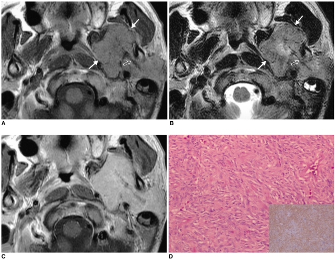
We retrospectively reviewed six MR images and two CT images of six histologically proven cases of SFT that occurred in four men and two women, and their ages ranged from 46 to 59 years. These imaging findings were correlated with the microscopic findings of their surgical specimens.

RESULTS

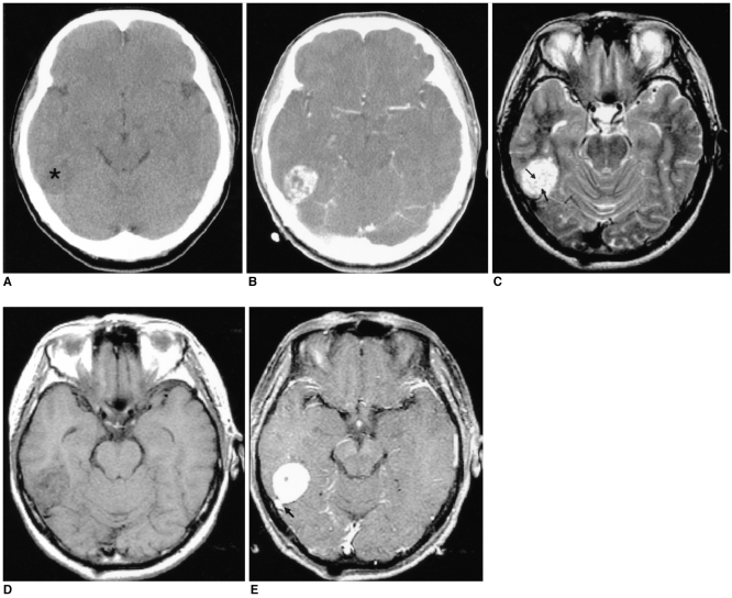
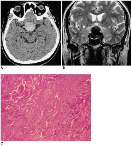
Six SFTs arose in the meninges (the petrous ridge and the pituitary fossa), the parotid gland, the parapharyngeal space, the buccal space and the maxillary sinus. On the MR images, SFTs in the intracranial and extracranial head and neck regions were mostly isointense to the muscle on the T1-weighted images, they were hyperintense on the T2-weighted images and they all had intense enhancement. On the T1- and T2-weighted images, hypointense lines were observed within in five SFTs. On the CT images, the SFTs were hypodense to the muscle on the unenhanced images and they were heterogeneously enhanced on the contrast-enhanced images. An exceptional case of pituitary SFT was hypointense on the T2-weighted images and it was hyperdense on the unenhanced CT images, which correlated with the increased collagenous component and the cellular compactness.

CONCLUSION

The imaging features of SFT are nonspecific; however, SFT should be included in the differential diagnosis of masses involving the intracranial and extracranial head and neck regions.

摘要

目的

孤立性纤维瘤(SFT)是一种非常罕见的肿瘤。本研究的目的是确定颅内及颅外头颈部区域SFT的磁共振成像(MR)特征。

材料与方法

我们回顾性分析了6例经组织学证实的SFT患者的6幅MR图像和2幅CT图像,其中4例男性,2例女性,年龄在46至59岁之间。这些影像学表现与手术标本的显微镜检查结果相关。

结果

6例SFT分别发生于脑膜(岩骨嵴和垂体窝)、腮腺、咽旁间隙、颊间隙和上颌窦。在MR图像上,颅内及颅外头颈部区域的SFT在T1加权图像上大多与肌肉等信号,在T2加权图像上呈高信号,且均有明显强化。在T1加权和T2加权图像上,5例SFT内可见低信号线。在CT图像上,SFT在平扫图像上相对于肌肉呈低密度,在增强图像上呈不均匀强化。1例垂体SFT在T2加权图像上呈低信号,在平扫CT图像上呈高密度,这与胶原成分增加和细胞致密性增加有关。

结论

SFT的影像学表现无特异性;然而,在涉及颅内及颅外头颈部区域肿块的鉴别诊断中应考虑SFT。

https://cdn.ncbi.nlm.nih.gov/pmc/blobs/45c8/2685036/2bdc2ef04e68/kjr-6-136-g003.jpg
https://cdn.ncbi.nlm.nih.gov/pmc/blobs/45c8/2685036/4c9f83845ad7/kjr-6-136-g001.jpg
https://cdn.ncbi.nlm.nih.gov/pmc/blobs/45c8/2685036/6cca762135d7/kjr-6-136-g002.jpg
https://cdn.ncbi.nlm.nih.gov/pmc/blobs/45c8/2685036/2bdc2ef04e68/kjr-6-136-g003.jpg
https://cdn.ncbi.nlm.nih.gov/pmc/blobs/45c8/2685036/4c9f83845ad7/kjr-6-136-g001.jpg
https://cdn.ncbi.nlm.nih.gov/pmc/blobs/45c8/2685036/6cca762135d7/kjr-6-136-g002.jpg
https://cdn.ncbi.nlm.nih.gov/pmc/blobs/45c8/2685036/2bdc2ef04e68/kjr-6-136-g003.jpg

相似文献

1
MR imaging of solitary fibrous tumors in the head and neck.头颈部孤立性纤维瘤的磁共振成像
Korean J Radiol. 2005 Jul-Sep;6(3):136-42. doi: 10.3348/kjr.2005.6.3.136.
2
Solitary fibrous tumours in the extracranial head and neck region: correlation of CT and MR features with pathologic findings.颅外头颈部区域的孤立性纤维性肿瘤:CT和MR特征与病理结果的相关性
Radiol Med. 2014 Dec;119(12):910-919. doi: 10.1007/s11547-014-0409-9. Epub 2014 May 27.
3
Computed tomography and magnetic resonance imaging findings of solitary fibrous tumors in the pelvis: correlation with histopathological findings.骨盆孤立性纤维瘤的计算机断层扫描和磁共振成像表现:与组织病理学发现的相关性。
Eur J Radiol. 2011 Apr;78(1):65-70. doi: 10.1016/j.ejrad.2009.09.001. Epub 2009 Oct 7.
4
Computed tomography and magnetic resonance imaging findings of inflammatory myofibroblastic tumors of the head and neck.头颈部炎性肌纤维母细胞瘤的计算机断层扫描和磁共振成像表现
Acta Radiol. 2014 May;55(4):434-40. doi: 10.1177/0284185113500165. Epub 2013 Aug 21.
5
Computed tomography and magnetic resonance imaging findings of peripheral primitive neuroectodermal tumors of the head and neck.头颈部外周原始神经外胚层肿瘤的计算机断层扫描和磁共振成像表现。
Eur J Radiol. 2011 Nov;80(2):607-11. doi: 10.1016/j.ejrad.2011.02.008. Epub 2011 Feb 26.
6
Solitary fibrous tumors of the central nervous system: clinical features and imaging findings in 22 patients.中枢神经系统孤立性纤维瘤:22例患者的临床特征及影像学表现
J Comput Assist Tomogr. 2013 Sep-Oct;37(5):658-65. doi: 10.1097/RCT.0b013e3182a05687.
7
Solitary fibrous tumor of the orbit: CT and MR imaging findings.眼眶孤立性纤维瘤:CT与MR成像表现
AJNR Am J Neuroradiol. 2008 May;29(5):857-62. doi: 10.3174/ajnr.A0961. Epub 2008 Feb 13.
8
Head and neck solitary fibrous tumors: a rare and challenging entity.头颈部孤立性纤维瘤:一种罕见且具有挑战性的疾病。
Eur Arch Otorhinolaryngol. 2016 Jun;273(6):1589-98. doi: 10.1007/s00405-015-3670-1. Epub 2015 May 31.
9
Solitary fibrous tumor of the sinonasal cavity: CT and MR imaging findings.鼻腔鼻窦孤立性纤维瘤:CT 和 MR 成像表现。
AJNR Am J Neuroradiol. 2013 Jun-Jul;34(6):1248-51. doi: 10.3174/ajnr.A3485. Epub 2013 Feb 14.
10
MRI study of solitary fibrous tumor in the orbit.眼眶孤立性纤维瘤的 MRI 研究。
AJR Am J Roentgenol. 2012 Oct;199(4):W506-11. doi: 10.2214/AJR.11.8477.

引用本文的文献

1
Solitary fibrous tumor in the pineal region: A series of 5 cases and literature review.松果体区孤立性纤维瘤:5例病例系列及文献复习
J Neuropathol Exp Neurol. 2025 May 1;84(5):379-390. doi: 10.1093/jnen/nlaf008.
2
Solitary fibrous tumor of the adrenal gland: a case report and review of the literature.肾上腺孤立性纤维瘤:一例报告并文献复习
Front Surg. 2024 Jun 5;11:1363807. doi: 10.3389/fsurg.2024.1363807. eCollection 2024.
3
Freiburg Neuropathology Case Conference : A 43-year-old Patient Presenting with a Right-sided, Slowly Progressing, Painless Exophthalmos and Ptosis.

本文引用的文献

1
Solitary fibrous tumors of the pleura: surgical outcome and clinical course.胸膜孤立性纤维瘤:手术结果与临床病程
Ann Thorac Surg. 2005 Jan;79(1):303-7. doi: 10.1016/j.athoracsur.2004.07.013.
2
Solitary fibrous tumor of the spinal cord: a clinicopathologic study of two cases.脊髓孤立性纤维瘤:两例临床病理研究
Ann Diagn Pathol. 2004 Oct;8(5):268-75. doi: 10.1016/j.anndiagpath.2004.07.002.
3
Solitary fibrous tumor of the parapharyngeal space: MR imaging findings.咽旁间隙孤立性纤维瘤:磁共振成像表现
弗莱堡神经病理学病例研讨会:一名43岁患者,表现为右侧、缓慢进展、无痛性眼球突出和上睑下垂。
Clin Neuroradiol. 2023 Mar;33(1):255-260. doi: 10.1007/s00062-023-01267-1. Epub 2023 Feb 20.
4
Magnetic resonance imaging features of a solitary fibrous tumor of the vulva: a case report.外阴孤立性纤维瘤的磁共振成像特征:一例报告。
J Int Med Res. 2022 Jul;50(7):3000605221112201. doi: 10.1177/03000605221112201.
5
Parotid Gland Solitary Fibrous Tumor Presenting as a Long Duration Mass: A Case Report.以长期肿块为表现的腮腺孤立性纤维瘤:一例报告
Case Rep Pathol. 2022 Feb 24;2022:2097634. doi: 10.1155/2022/2097634. eCollection 2022.
6
Solitary Fibrous Tumor or Hemangiopericytoma of the Sella in an Older Patient Treated with Partial Removal Followed by Fractionated Gamma Knife Radiosurgery.老年患者蝶鞍区孤立性纤维瘤或血管外皮细胞瘤经部分切除后行分次伽玛刀放射外科治疗
NMC Case Rep J. 2021 Oct 16;8(1):697-703. doi: 10.2176/nmccrj.cr.2021-0103. eCollection 2021.
7
Endoscopic resection of solitary fibrous tumor of the ethmoid: Case report review of the literature.筛窦孤立性纤维瘤的内镜下切除术:病例报告及文献复习
Ann Med Surg (Lond). 2020 Dec 7;60:714-718. doi: 10.1016/j.amsu.2020.11.073. eCollection 2020 Dec.
8
Rapidly growing giant solitary fibrous tumor of the pleura: a case report and review of the literature.迅速生长的胸膜巨大孤立性纤维瘤:一例报告并文献复习
Int J Clin Exp Pathol. 2020 Sep 1;13(9):2363-2368. eCollection 2020.
9
Solitary fibrous tumour of the parotid gland: a case report and a 15-year literature review.腮腺孤立性纤维瘤:一例报告及15年文献综述
AME Case Rep. 2019 May 24;3:14. doi: 10.21037/acr.2019.04.05. eCollection 2019.
10
A series of two cases of rare tumors: Solitary fibrous tumor of the pleura.两例罕见肿瘤病例系列:胸膜孤立性纤维瘤。
Respir Med Case Rep. 2019 Jun 4;28:100872. doi: 10.1016/j.rmcr.2019.100872. eCollection 2019.
AJNR Am J Neuroradiol. 2002 Mar;23(3):473-5.
4
Solitary fibrous tumor of the pleura: MR appearance and enhancement pattern.胸膜孤立性纤维瘤:磁共振成像表现及强化方式
J Comput Assist Tomogr. 2002 Mar-Apr;26(2):174-9. doi: 10.1097/00004728-200203000-00002.
5
Solitary fibrous tumours of the meninges: case series and literature review.脑膜孤立性纤维瘤:病例系列及文献综述
J Neurooncol. 2001 Aug;54(1):57-69. doi: 10.1023/a:1012553119349.
6
Solitary fibrous tumor in the buccal space: MR findings with pathologic correlation.颊间隙孤立性纤维瘤:磁共振成像表现与病理对照
AJNR Am J Neuroradiol. 2001 Nov-Dec;22(10):1890-2.
7
Solitary fibrous tumor of the nasal cavity.鼻腔孤立性纤维瘤
Otolaryngol Head Neck Surg. 1999 Dec;121(6):826-8. doi: 10.1053/hn.1999.v121.a95230.
8
Solitary fibrous tumor of the parotid gland extending to the parapharyngeal space.腮腺孤立性纤维瘤延伸至咽旁间隙。
Eur Arch Otorhinolaryngol. 1998;255(1):18-21. doi: 10.1007/s004050050015.
9
Localized benign fibrous tumors of the pleura: MR appearance.胸膜局限性良性纤维瘤:磁共振成像表现
J Comput Assist Tomogr. 1997 Jan-Feb;21(1):115-20. doi: 10.1097/00004728-199701000-00023.
10
Solitary fibrous tumor of the paranasal sinuses: CT and MR appearance.鼻窦孤立性纤维瘤:CT与MR表现
AJNR Am J Neuroradiol. 1996 Oct;17(9):1767-72.