• 文献检索
  • 文档翻译
  • 深度研究
  • 学术资讯
  • Suppr Zotero 插件Zotero 插件
  • 邀请有礼
  • 套餐&价格
  • 历史记录
应用&插件
Suppr Zotero 插件Zotero 插件浏览器插件Mac 客户端Windows 客户端微信小程序
定价
高级版会员购买积分包购买API积分包
服务
文献检索文档翻译深度研究API 文档MCP 服务
关于我们
关于 Suppr公司介绍联系我们用户协议隐私条款
关注我们

Suppr 超能文献

核心技术专利:CN118964589B侵权必究
粤ICP备2023148730 号-1Suppr @ 2026

文献检索

告别复杂PubMed语法,用中文像聊天一样搜索,搜遍4000万医学文献。AI智能推荐,让科研检索更轻松。

立即免费搜索

文件翻译

保留排版,准确专业,支持PDF/Word/PPT等文件格式,支持 12+语言互译。

免费翻译文档

深度研究

AI帮你快速写综述,25分钟生成高质量综述,智能提取关键信息,辅助科研写作。

立即免费体验

X线、计算机断层扫描及磁共振成像在22例累及长骨的儿童朗格汉斯细胞组织细胞增多症患者中的应用:一项回顾性分析。

The application of x-ray, computed tomography, and magnetic resonance imaging on 22 pediatric Langerhans cell histiocytosis patients with long bone involvement: A retrospective analysis.

作者信息

Zhang Xiaojun, Zhou Jing, Chai Xuee, Chen Guiling, Guo Bin, Ni Lei, Wu Peng

机构信息

Department of Radiology.

Department of Radiology, Affiliated Hospital of Nanjing University of Chinese Medicine.

出版信息

Medicine (Baltimore). 2018 Apr;97(17):e0411. doi: 10.1097/MD.0000000000010411.

DOI:10.1097/MD.0000000000010411
PMID:29702989
原文链接:https://pmc.ncbi.nlm.nih.gov/articles/PMC5944563/
Abstract

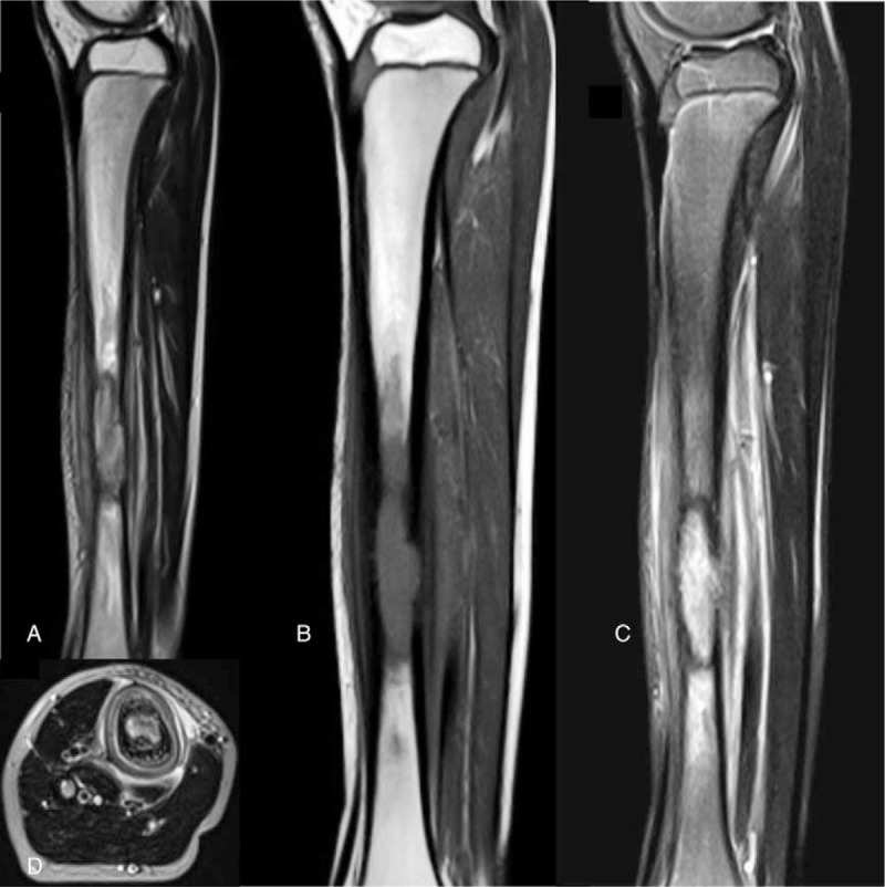
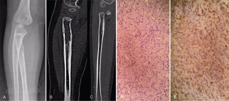
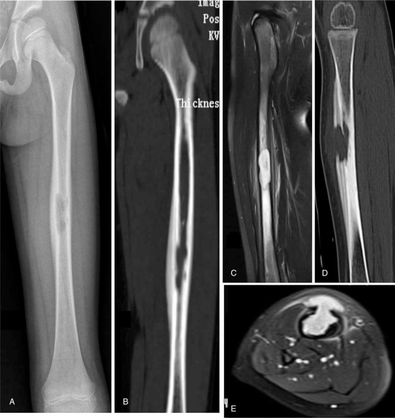
The studies focusing on x-ray, computed tomography (CT), and magnetic resonance imaging (MRI) in pediatric Langerhans cell histiocytosis (LCH) patients were still rare. Therefore, we aimed to evaluate the application of x-ray, CT, and MRI in pediatric LCH patients with long bone involvement.Total 22 pediatric LCH patients were included in this study. The diagnosis of LCH was confirmed by pathological examination. All patients were followed up for 3 years. X-ray, CT, or MRI was performed and the results were recorded for further analyses.Among 22 pediatric patients, x-ray (n = 20), CT (n = 18), or MRI (n = 12) were used to scan the lesion on long bones affected by LCH. Femurs (n = 13, 38.24%), tibia (n = 11, 32.35%), humerus (n = 5, 14.71%), and radius (n = 4, 11.76%) were the most frequently affected anatomic sites. Ovoid or round radiolucent lesions, aggressive periosteal reaction, and swelling of surrounding soft tissues were characteristic image of long bones on x-ray, CT, and MRI in pediatric LCH.Femurs, tibia, humerus, and radius were the most commonly affected long bones of pediatric LCH. The application of x-ray, CT, and MRI on long bones could help with the diagnosis of pediatric LCH.

摘要

针对小儿朗格汉斯细胞组织细胞增生症(LCH)患者的X线、计算机断层扫描(CT)和磁共振成像(MRI)的研究仍然很少。因此,我们旨在评估X线、CT和MRI在患有长骨受累的小儿LCH患者中的应用。本研究共纳入22例小儿LCH患者。LCH的诊断经病理检查确诊。所有患者均随访3年。进行了X线、CT或MRI检查,并记录结果以供进一步分析。在22例小儿患者中,使用X线(n = 20)、CT(n = 18)或MRI(n = 12)扫描受LCH影响的长骨病变。股骨(n = 13,38.24%)、胫骨(n = 11,32.35%)、肱骨(n = 5,14.71%)和桡骨(n = 4,11.76%)是最常受累的解剖部位。椭圆形或圆形透亮病变、侵袭性骨膜反应以及周围软组织肿胀是小儿LCH患者长骨在X线、CT和MRI上的特征性影像。股骨、胫骨、肱骨和桡骨是小儿LCH最常受累的长骨。X线、CT和MRI在长骨上的应用有助于小儿LCH的诊断。

https://cdn.ncbi.nlm.nih.gov/pmc/blobs/0c07/5944563/2c7bb039ba68/medi-97-e0411-g005.jpg
https://cdn.ncbi.nlm.nih.gov/pmc/blobs/0c07/5944563/5434a6fd6b82/medi-97-e0411-g002.jpg
https://cdn.ncbi.nlm.nih.gov/pmc/blobs/0c07/5944563/49fb666d57fd/medi-97-e0411-g003.jpg
https://cdn.ncbi.nlm.nih.gov/pmc/blobs/0c07/5944563/2c7bb039ba68/medi-97-e0411-g005.jpg
https://cdn.ncbi.nlm.nih.gov/pmc/blobs/0c07/5944563/5434a6fd6b82/medi-97-e0411-g002.jpg
https://cdn.ncbi.nlm.nih.gov/pmc/blobs/0c07/5944563/49fb666d57fd/medi-97-e0411-g003.jpg
https://cdn.ncbi.nlm.nih.gov/pmc/blobs/0c07/5944563/2c7bb039ba68/medi-97-e0411-g005.jpg

相似文献

1
The application of x-ray, computed tomography, and magnetic resonance imaging on 22 pediatric Langerhans cell histiocytosis patients with long bone involvement: A retrospective analysis.X线、计算机断层扫描及磁共振成像在22例累及长骨的儿童朗格汉斯细胞组织细胞增多症患者中的应用:一项回顾性分析。
Medicine (Baltimore). 2018 Apr;97(17):e0411. doi: 10.1097/MD.0000000000010411.
2
MRI and clinical features of Langerhans cell histiocytosis (LCH) in the pelvis and extremities: can LCH really look like anything?骨盆和四肢朗格汉斯细胞组织细胞增多症(LCH)的MRI及临床特征:LCH真的可以表现出任何样子吗?
Skeletal Radiol. 2016 May;45(5):607-13. doi: 10.1007/s00256-016-2330-x. Epub 2016 Jan 22.
3
Clear cell chondrosarcoma: radiographic, computed tomographic, and magnetic resonance findings in 34 patients with pathologic correlation.透明细胞软骨肉瘤:34例患者的X线、计算机断层扫描及磁共振成像表现与病理对照
Skeletal Radiol. 2003 Dec;32(12):687-94. doi: 10.1007/s00256-003-0668-3. Epub 2003 Oct 7.
4
Solid variant of aneurysmal bone cysts in long tubular bones: giant cell reparative granuloma.长管状骨动脉瘤样骨囊肿的实性变体:巨细胞修复性肉芽肿。
AJR Am J Roentgenol. 2003 Jun;180(6):1681-7. doi: 10.2214/ajr.180.6.1801681.
5
Sonographic diagnosis and monitoring of localized langerhans cell histiocytosis of the skull.颅骨局限性朗格汉斯细胞组织细胞增多症的超声诊断与监测
J Clin Ultrasound. 2013 Mar-Apr;41(3):134-9. doi: 10.1002/jcu.21988. Epub 2012 Sep 18.
6
Langerhans cell histiocytosis of long bones: MR imaging and complete follow up study.长骨朗格汉斯细胞组织细胞增多症:磁共振成像及完整随访研究
J Cancer Res Ther. 2012 Apr-Jun;8(2):286-8. doi: 10.4103/0973-1482.98991.
7
Pediatric Langerhans cell histiocytosis of the temporal bone: clinical and imaging studies of 27 cases.儿童颞骨朗格汉斯细胞组织细胞增生症:27 例临床和影像学研究。
World J Surg Oncol. 2018 Mar 27;16(1):72. doi: 10.1186/s12957-018-1366-x.
8
Langerhans Cell Histiocytosis of the Temporal Bone with Otic Capsule Involvement.累及听骨囊的颞骨朗格汉斯细胞组织细胞增多症。
Clin Neuroradiol. 2017 Jun;27(2):163-168. doi: 10.1007/s00062-015-0461-4. Epub 2015 Sep 4.
9
[Clinical aspects and radiology of histiocytosis X in the skeleton--a retrospective study of 18 patients].[骨骼嗜酸性肉芽肿的临床与放射学表现——18例患者的回顾性研究]
Rontgenblatter. 1985 Dec;38(12):400-6.
10
[Bone scintigraphy and somatostatin receptor scintigraphy in pediatric patients with bone involvement in Langerhans cell histiocytosis].[骨闪烁显像和生长抑素受体闪烁显像在朗格汉斯细胞组织细胞增多症骨受累儿科患者中的应用]
Rev Esp Med Nucl. 2003 Nov;22(6):367-75. doi: 10.1016/s0212-6982(03)72220-4.

引用本文的文献

1
Characteristics and radiological features of bone lesions in patients with Langerhans cell histiocytosis: A case series study.朗格汉斯细胞组织细胞增多症患者骨病变的特征及影像学表现:一项病例系列研究。
Medicine (Baltimore). 2025 Mar 14;104(11):e41833. doi: 10.1097/MD.0000000000041833.
2
Infantile BCG osteomyelitis ultimately diagnosed as a result of trauma.婴儿卡介苗骨髓炎最终因创伤而被诊断出来。
Radiol Case Rep. 2025 Jan 13;20(3):1784-1789. doi: 10.1016/j.radcr.2024.12.034. eCollection 2025 Mar.
3
Dural involvement in central nervous system langerhans cells histiocytosis (LCH) on FDG PET/CT: Case report and review of CNS manifestations of LCH on PET/CT.

本文引用的文献

1
Extraosseous Ewing Sarcoma: Diagnosis, Prognosis and Optimal Management.骨外尤文肉瘤:诊断、预后及最佳治疗
Indian J Surg. 2016 Feb;78(1):49-53. doi: 10.1007/s12262-015-1399-0. Epub 2015 Nov 13.
2
Langerhans Cell Histiocytosis: Emerging Insights and Clinical Implications.朗格汉斯细胞组织细胞增多症:新见解与临床意义
Oncology (Williston Park). 2016 Feb;30(2):122-32, 139.
3
Langerhans Cell Histiocytosis: A Clinicopathologic Review and Molecular Pathogenetic Update.朗格汉斯细胞组织细胞增多症:临床病理回顾与分子发病机制更新
18F-FDG PET/CT显示硬脑膜受累的中枢神经系统朗格汉斯细胞组织细胞增多症(LCH):病例报告及PET/CT上LCH中枢神经系统表现的文献复习
Radiol Case Rep. 2024 Jan 15;19(4):1391-1396. doi: 10.1016/j.radcr.2023.12.044. eCollection 2024 Apr.
4
The baseline metabolism parameters of F‑FDG PET/CT as promising prognostic biomarkers in pediatric Langerhans cell histiocytosis.F-FDG PET/CT的基线代谢参数作为儿童朗格汉斯细胞组织细胞增多症中有前景的预后生物标志物。
Quant Imaging Med Surg. 2023 Sep 1;13(9):5934-5944. doi: 10.21037/qims-23-290. Epub 2023 Aug 4.
5
Clinical characteristics and outcomes of Langerhans cell histiocytosis at a single institution in Thailand: a 20-year retrospective study.泰国一家机构朗格汉斯细胞组织细胞增多症的临床特征与结局:一项20年回顾性研究
Asian Biomed (Res Rev News). 2021 Aug 20;15(4):171-181. doi: 10.2478/abm-2021-0022. eCollection 2021 Aug.
6
Role of F-FDG PET/CT in the diagnosis and management of patients with Langerhans cell histiocytosis.氟代脱氧葡萄糖正电子发射断层扫描/计算机断层扫描(F-FDG PET/CT)在朗格汉斯细胞组织细胞增多症患者诊断及管理中的作用
Quant Imaging Med Surg. 2022 Jun;12(6):3351-3363. doi: 10.21037/qims-21-823.
7
F-FDG PET/MR Assessment of Pediatric Langerhans Cell Histiocytosis.小儿朗格汉斯细胞组织细胞增多症的F-FDG PET/MR评估
Int J Gen Med. 2021 Oct 1;14:6251-6259. doi: 10.2147/IJGM.S327134. eCollection 2021.
8
Multisystem Langerhans Cell Histiocytosis in an infant.一名婴儿的多系统朗格汉斯细胞组织细胞增多症
Radiol Case Rep. 2021 May 6;16(7):1798-1805. doi: 10.1016/j.radcr.2021.04.028. eCollection 2021 Jul.
9
Radiologic findings that aid in the reduction of misdiagnoses of Langerhans cell histiocytosis of the bone: a retrospective study.有助于降低骨朗格汉斯细胞组织细胞增生症误诊的放射学表现:一项回顾性研究。
World J Surg Oncol. 2021 May 10;19(1):146. doi: 10.1186/s12957-021-02261-y.
10
Langerhans cell histiocytosis of the jaw, a mimicker of osteomyelitis on CT and MR images: A retrospective analysis.颌骨朗格汉斯细胞组织细胞增多症:CT和MR图像上酷似骨髓炎——一项回顾性分析
Medicine (Baltimore). 2019 Jul;98(27):e16331. doi: 10.1097/MD.0000000000016331.
Arch Pathol Lab Med. 2015 Oct;139(10):1211-4. doi: 10.5858/arpa.2015-0199-RA.
4
Dendritic Cells Cause Bone Lesions in a New Mouse Model of Histiocytosis.树突状细胞在一种新的组织细胞增多症小鼠模型中引发骨病变。
PLoS One. 2015 Aug 6;10(8):e0133917. doi: 10.1371/journal.pone.0133917. eCollection 2015.
5
S3-Guideline on allergy prevention: 2014 update: Guideline of the German Society for Allergology and Clinical Immunology (DGAKI) and the German Society for Pediatric and Adolescent Medicine (DGKJ).《过敏预防S3指南:2014年更新版》:德国变态反应与临床免疫学会(DGAKI)及德国儿科与青少年医学学会(DGKJ)指南
Allergo J Int. 2014;23(6):186-199. doi: 10.1007/s40629-014-0022-4.
6
Pulmonary Langerhans Cell Histiocytosis with Lytic Bone Involvement in an Adult Smoker: Regression following Smoking Cessation.成年吸烟者合并溶骨性骨受累的肺朗格汉斯细胞组织细胞增多症:戒烟后病情缓解
Case Rep Hematol. 2015;2015:201536. doi: 10.1155/2015/201536. Epub 2015 Feb 18.
7
Langerhans cell histiocytosis.朗格汉斯细胞组织细胞增多症
Cancer Control. 2014 Oct;21(4):328-34. doi: 10.1177/107327481402100409.
8
Diagnosis and management of langerhans cell histiocytosis.朗格汉斯细胞组织细胞增多症的诊断与管理
J Am Acad Orthop Surg. 2014 Oct;22(10):643-52. doi: 10.5435/JAAOS-22-10-643.
9
Recent advances in Langerhans cell histiocytosis.朗格汉斯细胞组织细胞增多症的最新进展
Pediatr Int. 2014 Aug;56(4):451-61. doi: 10.1111/ped.12380.
10
Langerhans cell histiocytosis of the clavicle in a 13-year-old boy.一名13岁男孩的锁骨朗格汉斯细胞组织细胞增多症。
Case Rep Orthop. 2014;2014:510287. doi: 10.1155/2014/510287. Epub 2014 Jan 5.