• 文献检索
  • 文档翻译
  • 深度研究
  • 学术资讯
  • Suppr Zotero 插件Zotero 插件
  • 邀请有礼
  • 套餐&价格
  • 历史记录
应用&插件
Suppr Zotero 插件Zotero 插件浏览器插件Mac 客户端Windows 客户端微信小程序
定价
高级版会员购买积分包购买API积分包
服务
文献检索文档翻译深度研究API 文档MCP 服务
关于我们
关于 Suppr公司介绍联系我们用户协议隐私条款
关注我们

Suppr 超能文献

核心技术专利:CN118964589B侵权必究
粤ICP备2023148730 号-1Suppr @ 2026

文献检索

告别复杂PubMed语法,用中文像聊天一样搜索,搜遍4000万医学文献。AI智能推荐,让科研检索更轻松。

立即免费搜索

文件翻译

保留排版,准确专业,支持PDF/Word/PPT等文件格式,支持 12+语言互译。

免费翻译文档

深度研究

AI帮你快速写综述,25分钟生成高质量综述,智能提取关键信息,辅助科研写作。

立即免费体验

使用三维T2加权稳态自由进动序列对球囊进行成像以诊断梅尼埃病中的内淋巴积水:准确、快速且无需静脉注射造影剂。

Imaging of the saccule for the diagnosis of endolymphatic hydrops in Meniere disease, using a three-dimensional T2-weighted steady state free precession sequence: accurate, fast, and without contrast material intravenous injection.

作者信息

Venkatasamy Aïna, Veillon Francis, Fleury Aude, Eliezer Michael, Abu Eid Maher, Romain Benoit, Vuong Hella, Rohmer Dominique, Charpiot Anne, Sick Henri, Riehm Sophie

机构信息

1Service d'imagerie 1, Hôpitaux Universitaires de Strasbourg, 1 avenue Molière, Strasbourg, F-67098 France.

2Service d'ORL, Hôpitaux Universitaires de Strasbourg, Strasbourg, France.

出版信息

Eur Radiol Exp. 2017;1(1):14. doi: 10.1186/s41747-017-0020-7. Epub 2017 Oct 9.

DOI:10.1186/s41747-017-0020-7
PMID:29708183
原文链接:https://pmc.ncbi.nlm.nih.gov/articles/PMC5909345/
Abstract

BACKGROUND

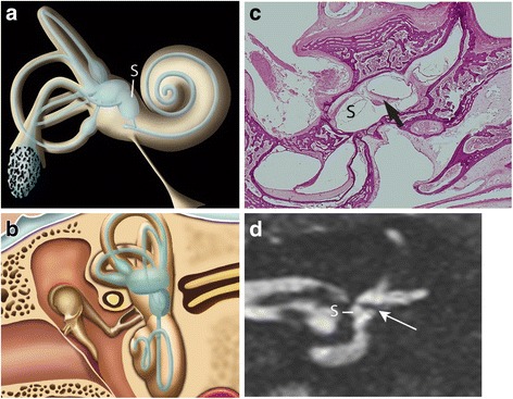
Endolymphatic hydrops can be studied on magnetic resonance imaging (MRI) using images acquired 4 h after intravenous injection of Gd-chelate. Our aim was to compare high-resolution T2-weighted images of the saccule in normal subjects with histological sections from cadavers and to identify its changes in Meniere disease, compared to healthy volunteers.

METHODS

Sixty-four healthy volunteers without any otologic disease and 64 patients who fulfilled all the criteria for unilateral Meniere disease underwent 3 T MRI using a T2-weighted steady state free precession (SSFP) sequence, without contrast material injection. Images of healthy volunteers were compared with histological sections of normal inner ears from premature foetuses and compared with volunteers.

RESULTS

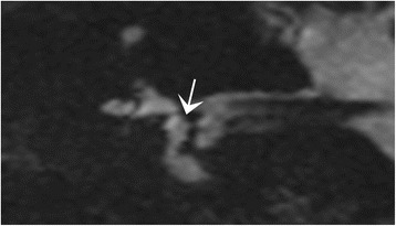
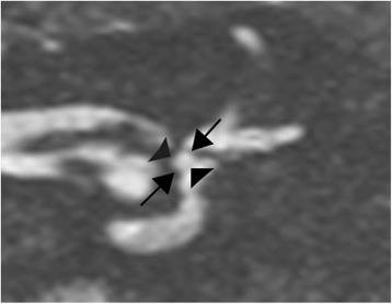
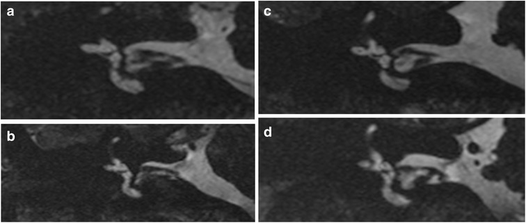
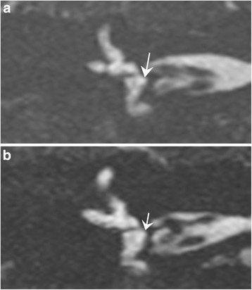
The normal saccule was easily visible on T2-weighted images in volunteers, with a normal maximal height of 1.6 mm (1.4 ± 0.1 mm, mean ± standard deviation) and a good correlation with reference histological sections, while in Meniere disease the saccule was dilated in 52/62 patients (84%), with a saccular height greater than 1.6 mm (1.69 ± 0.24 mm,  = 0.001), found in 45/52 patients (86%). An associated increased width (greater than 1.4 mm) was found in 23/52 patients (44%). A round shape or the non-visualisation of the saccule were also found in 2/52 (4%) and in 5/62 patients (8%), respectively.

CONCLUSIONS

A T2-weighted sequence is an easy method to diagnose Meniere disease. Saccular abnormalities were found in 84% of the cases: elongation (height > 1.6 mm) in 86%, increased saccular width in 44%, or a missing saccule in 8%.

摘要

背景

内淋巴积水可通过静脉注射钆螯合物4小时后采集的磁共振成像(MRI)进行研究。我们的目的是将正常受试者球囊的高分辨率T2加权图像与尸体组织切片进行比较,并确定与健康志愿者相比,梅尼埃病患者球囊的变化。

方法

64名无任何耳科疾病的健康志愿者和64名符合单侧梅尼埃病所有标准的患者接受了3T MRI检查,使用T2加权稳态自由进动(SSFP)序列,未注射造影剂。将健康志愿者的图像与早产胎儿正常内耳的组织切片进行比较,并与志愿者进行比较。

结果

在志愿者的T2加权图像上,正常球囊很容易看到,正常最大高度为1.6毫米(1.4±0.1毫米,平均值±标准差)与参考组织切片有良好的相关性,而在梅尼埃病中,52/62例患者(84%)的球囊扩张,球囊高度大于1.6毫米(1.69±0.24毫米,P=0.001),45/52例患者(86%)出现这种情况;23/52例患者(44%)发现球囊宽度增加(大于1.4毫米);2/52例(4%)和5/62例患者(8%)分别出现球囊呈圆形或未显示的情况。

结论

T2加权序列是诊断梅尼埃病的简便方法。84%的病例发现球囊异常:86%为伸长(高度>1.6毫米),44%为球囊宽度增加,8%为球囊缺失。

https://cdn.ncbi.nlm.nih.gov/pmc/blobs/a134/6091711/4f8e62d18fb3/41747_2017_20_Fig6_HTML.jpg
https://cdn.ncbi.nlm.nih.gov/pmc/blobs/a134/6091711/688ad0e309fa/41747_2017_20_Fig1_HTML.jpg
https://cdn.ncbi.nlm.nih.gov/pmc/blobs/a134/6091711/c7517baea700/41747_2017_20_Fig2_HTML.jpg
https://cdn.ncbi.nlm.nih.gov/pmc/blobs/a134/6091711/645a94321668/41747_2017_20_Fig3_HTML.jpg
https://cdn.ncbi.nlm.nih.gov/pmc/blobs/a134/6091711/7927a53c1153/41747_2017_20_Fig4_HTML.jpg
https://cdn.ncbi.nlm.nih.gov/pmc/blobs/a134/6091711/72bd0f0a5bbb/41747_2017_20_Fig5_HTML.jpg
https://cdn.ncbi.nlm.nih.gov/pmc/blobs/a134/6091711/4f8e62d18fb3/41747_2017_20_Fig6_HTML.jpg

相似文献

1
Imaging of the saccule for the diagnosis of endolymphatic hydrops in Meniere disease, using a three-dimensional T2-weighted steady state free precession sequence: accurate, fast, and without contrast material intravenous injection.使用三维T2加权稳态自由进动序列对球囊进行成像以诊断梅尼埃病中的内淋巴积水:准确、快速且无需静脉注射造影剂。
Eur Radiol Exp. 2017;1(1):14. doi: 10.1186/s41747-017-0020-7. Epub 2017 Oct 9.
2
Saccular measurements in routine MRI can predict hydrops in Menière's disease.常规MRI中的囊状测量可预测梅尼埃病中的积水情况。
Eur Arch Otorhinolaryngol. 2017 Dec;274(12):4113-4120. doi: 10.1007/s00405-017-4756-8. Epub 2017 Sep 26.
3
MRI of endolymphatic hydrops in patients with intralabyrinthine schwannomas: a case-controlled study using non-enhanced T2-weighted images at 3 T.MRI 对前庭神经鞘瘤患者内淋巴积水的诊断:一项使用 3T 非增强 T2 加权成像的病例对照研究。
Eur Arch Otorhinolaryngol. 2021 Jun;278(6):1821-1827. doi: 10.1007/s00405-020-06271-6. Epub 2020 Aug 8.
4
Visualization of the saccule and utricle with non-contrast-enhanced FLAIR sequences.使用非增强 FLAIR 序列对球囊和椭圆囊进行可视化。
Eur Radiol. 2022 May;32(5):3532-3540. doi: 10.1007/s00330-021-08403-w. Epub 2021 Dec 20.
5
MRI of endolymphatic hydrops in patients with vestibular schwannomas: a case-controlled study using non-enhanced T2-weighted images at 3 Teslas.前庭神经鞘瘤患者内淋巴积水的 MRI:使用 3T 场强非增强 T2 加权成像的病例对照研究。
Eur Arch Otorhinolaryngol. 2019 Jun;276(6):1591-1599. doi: 10.1007/s00405-019-05395-8. Epub 2019 Mar 27.
6
Optimized 3D-FLAIR sequences to shorten the delay between intravenous administration of gadolinium and MRI acquisition in patients with Menière's disease.优化 3D-FLAIR 序列以缩短美尼尔氏病患者静脉内给予钆后至 MRI 采集的延迟时间。
Eur Radiol. 2022 Oct;32(10):6900-6909. doi: 10.1007/s00330-022-08889-y. Epub 2022 Jun 27.
7
MRI of endolymphatic hydrops in patients with Meniere's disease: a case-controlled study with a simplified classification based on saccular morphology.梅尼埃病患者内淋巴积水的磁共振成像:一项基于球囊形态学简化分类的病例对照研究
Eur Radiol. 2017 Aug;27(8):3138-3146. doi: 10.1007/s00330-016-4701-z. Epub 2016 Dec 20.
8
Membranous labyrinth volumes in normal ears and Ménière disease: a three-dimensional reconstruction study.正常耳与梅尼埃病的膜迷路容积:一项三维重建研究。
Laryngoscope. 2009 Nov;119(11):2216-20. doi: 10.1002/lary.20723.
9
The Correlation Between Endolymphatic Hydrops and Clinical Features of Meniere Disease.内淋巴积水与梅尼埃病临床特征的相关性。
Laryngoscope. 2021 Jan;131(1):E144-E150. doi: 10.1002/lary.28576. Epub 2020 Feb 21.
10
Relationship between video head impulse test, ocular and cervical vestibular evoked myogenic potentials, and compartmental magnetic resonance imaging classification in menière's disease.梅尼埃病中视频头脉冲试验、眼和颈前庭诱发肌源性电位与磁共振成像分区分类的关系。
Laryngoscope. 2020 Jul;130(7):E444-E452. doi: 10.1002/lary.28362. Epub 2019 Nov 19.

引用本文的文献

1
Utility and Challenges of Imaging in Peripheral Vestibular Disorder Diagnosis: A Narrative Review.影像学检查在外周性前庭疾病诊断中的作用与挑战:一篇叙述性综述
Diagnostics (Basel). 2025 May 16;15(10):1272. doi: 10.3390/diagnostics15101272.
2
Secondary endolymphatic hydrops: a clinical and literature overview.继发性内淋巴积水:临床与文献综述
Front Neurol. 2025 Feb 5;15:1525954. doi: 10.3389/fneur.2024.1525954. eCollection 2024.
3
A novel radiomics nomogram based on T2-sampling perfection with application-optimized contrasts using different flip-angle evolutions (SPACE) images for predicting cochlear and vestibular endolymphatic hydrops in Meniere's disease patients.

本文引用的文献

1
Differentiation of vestibular schwannomas from meningiomas of the internal auditory canal using perilymphatic signal evaluation on T2-weighted gradient-echo fast imaging employing steady state acquisition at 3T.在3T场强下,采用稳态采集的T2加权梯度回波快速成像,通过内淋巴信号评估区分前庭神经鞘瘤与内耳道脑膜瘤。
Eur Radiol Exp. 2017;1(1):8. doi: 10.1186/s41747-017-0012-7. Epub 2017 Jun 29.
2
Magnetic resonance imaging of intratympanic gadolinium helps differentiate vestibular migraine from Ménière disease.鼓室内钆的磁共振成像有助于鉴别前庭性偏头痛和梅尼埃病。
Laryngoscope. 2017 Oct;127(10):2382-2388. doi: 10.1002/lary.26518. Epub 2017 Feb 21.
3
一种基于 T2 采样完美的新型放射组学列线图,应用优化对比,使用不同翻转角演化(SPACE)图像,预测梅尼埃病患者耳蜗和前庭内淋巴积水。
Eur Radiol. 2024 Sep;34(9):6082-6091. doi: 10.1007/s00330-024-10670-2. Epub 2024 Mar 8.
4
pharmacological study of AQP2 inhibition by steroids contextualized to Ménière's disease treatments.针对梅尼埃病治疗,对类固醇抑制水通道蛋白2的药理学研究。
Front Neurol. 2023 Oct 19;14:1270092. doi: 10.3389/fneur.2023.1270092. eCollection 2023.
5
Magnetic resonance imaging of endolymphatic hydrops: a comparison of methods with and without gadolinium-based contrast agent administration.内淋巴积水的磁共振成像:有无钆基造影剂给药方法的比较。
Nagoya J Med Sci. 2023 May;85(2):299-309. doi: 10.18999/nagjms.85.2.299.
6
Confrontation of endolymphatic hydrops diagnosis on 3-Tesla MRI to clinical and audiovestibular findings in Meniere's disease.3特斯拉磁共振成像诊断内淋巴积水与梅尼埃病临床及听前庭检查结果的对比
Front Neurol. 2023 Jan 26;14:1105461. doi: 10.3389/fneur.2023.1105461. eCollection 2023.
7
Detailed insight into magnetic resonance assessment of Ménière's disease - description of methodology and imaging findings in a case series.梅尼埃病磁共振评估的深入见解——病例系列中的方法描述与影像学表现
Pol J Radiol. 2022 Jun 30;87:e354-e362. doi: 10.5114/pjr.2022.117971. eCollection 2022.
8
Editorial: Hydropic Ear Disease: Imaging and Functional Evaluation.社论:积水性耳病:影像学与功能评估
Front Surg. 2022 Apr 27;9:913741. doi: 10.3389/fsurg.2022.913741. eCollection 2022.
9
A Perspective for Ménière's Disease: In Silico Investigations of Dexamethasone as a Direct Modulator of AQP2.梅尼埃病的新视角:地塞米松作为 AQP2 直接调节剂的计算机模拟研究
Biomolecules. 2022 Mar 28;12(4):511. doi: 10.3390/biom12040511.
10
A non-invasive, automated diagnosis of Menière's disease using radiomics and machine learning on conventional magnetic resonance imaging: A multicentric, case-controlled feasibility study.基于常规磁共振成像的放射组学和机器学习对梅尼埃病进行非侵入性、自动化诊断:一项多中心、病例对照可行性研究。
Radiol Med. 2022 Jan;127(1):72-82. doi: 10.1007/s11547-021-01425-w. Epub 2021 Nov 25.
MRI of endolymphatic hydrops in patients with Meniere's disease: a case-controlled study with a simplified classification based on saccular morphology.
梅尼埃病患者内淋巴积水的磁共振成像:一项基于球囊形态学简化分类的病例对照研究
Eur Radiol. 2017 Aug;27(8):3138-3146. doi: 10.1007/s00330-016-4701-z. Epub 2016 Dec 20.
4
What is Menière's disease? A contemporary re-evaluation of endolymphatic hydrops.梅尼埃病是什么?对内淋巴积水的当代重新评估。
J Neurol. 2016 Apr;263 Suppl 1:S71-81. doi: 10.1007/s00415-015-7930-1. Epub 2016 Apr 15.
5
2015 Equilibrium Committee Amendment to the 1995 AAO-HNS Guidelines for the Definition of Ménière's Disease.2015年平衡委员会对1995年美国耳鼻咽喉头颈外科学会梅尼埃病定义指南的修订。
Otolaryngol Head Neck Surg. 2016 Mar;154(3):403-4. doi: 10.1177/0194599816628524. Epub 2016 Feb 16.
6
Diagnostic criteria for Menière's disease.梅尼埃病的诊断标准。
J Vestib Res. 2015;25(1):1-7. doi: 10.3233/VES-150549.
7
Endolymphatic hydrops and Ménière's disease: a lesion meta-analysis.内淋巴积水与梅尼埃病:一项病变荟萃分析。
J Laryngol Otol. 2014 Oct;128(10):859-65. doi: 10.1017/S0022215114001972. Epub 2014 Sep 19.
8
Visualization of endolymphatic hydrops with MR imaging in patients with Ménière's disease and related pathologies: current status of its methods and clinical significance.梅尼埃病及相关病变患者内淋巴积水的磁共振成像可视化:其方法的现状及临床意义
Jpn J Radiol. 2014 Apr;32(4):191-204. doi: 10.1007/s11604-014-0290-4. Epub 2014 Feb 6.
9
Decreased vestibular signal intensity on 3D-FIESTA in vestibular schwannomas differentiating from meningiomas.前庭神经鞘瘤的 3D-FIESTA 信号强度降低,有助于与脑膜瘤相鉴别。
Neuroradiology. 2013 Feb;55(3):261-70. doi: 10.1007/s00234-012-1100-2. Epub 2012 Oct 16.
10
Visualization of endolymphatic hydrops and correlation with audio-vestibular functional testing in patients with definite Meniere's disease.确诊梅尼埃病患者内淋巴积水的可视化及其与听-前庭功能测试的相关性。
Auris Nasus Larynx. 2013 Apr;40(2):167-72. doi: 10.1016/j.anl.2012.07.009. Epub 2012 Aug 4.