• 文献检索
  • 文档翻译
  • 深度研究
  • 学术资讯
  • Suppr Zotero 插件Zotero 插件
  • 邀请有礼
  • 套餐&价格
  • 历史记录
应用&插件
Suppr Zotero 插件Zotero 插件浏览器插件Mac 客户端Windows 客户端微信小程序
定价
高级版会员购买积分包购买API积分包
服务
文献检索文档翻译深度研究API 文档MCP 服务
关于我们
关于 Suppr公司介绍联系我们用户协议隐私条款
关注我们

Suppr 超能文献

核心技术专利:CN118964589B侵权必究
粤ICP备2023148730 号-1Suppr @ 2026

文献检索

告别复杂PubMed语法,用中文像聊天一样搜索,搜遍4000万医学文献。AI智能推荐,让科研检索更轻松。

立即免费搜索

文件翻译

保留排版,准确专业,支持PDF/Word/PPT等文件格式,支持 12+语言互译。

免费翻译文档

深度研究

AI帮你快速写综述,25分钟生成高质量综述,智能提取关键信息,辅助科研写作。

立即免费体验

碳离子放疗治疗复发性高级别胶质瘤中 F-FET PET 对靶区定义和肿瘤进展的影响。

Impact of F-FET PET on Target Volume Definition and Tumor Progression of Recurrent High Grade Glioma Treated with Carbon-Ion Radiotherapy.

机构信息

German Cancer Consortium (DKTK), Heidelberg, Germany.

Translational Radiation Oncology, National Center for Tumor Diseases (NCT), German Cancer Research Center (DKFZ), Heidelberg, Germany.

出版信息

Sci Rep. 2018 May 8;8(1):7201. doi: 10.1038/s41598-018-25350-7.

DOI:10.1038/s41598-018-25350-7
PMID:29740097
原文链接:https://pmc.ncbi.nlm.nih.gov/articles/PMC5940831/
Abstract

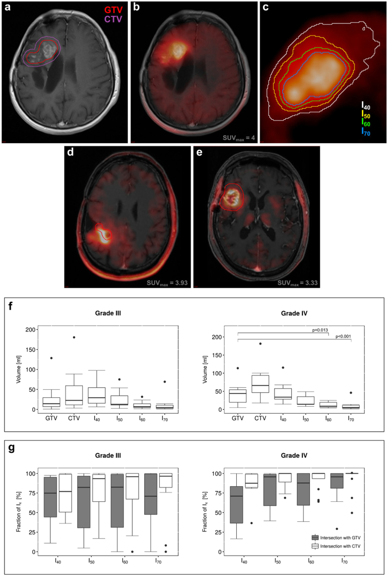
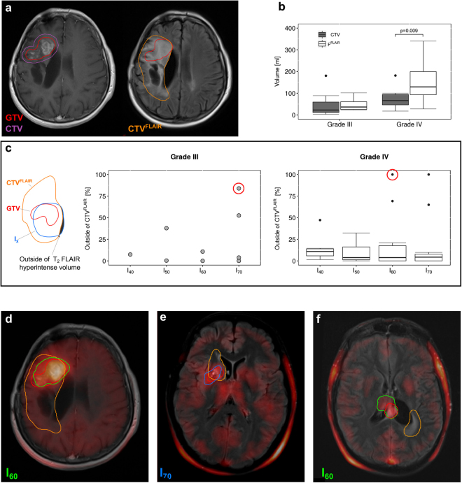
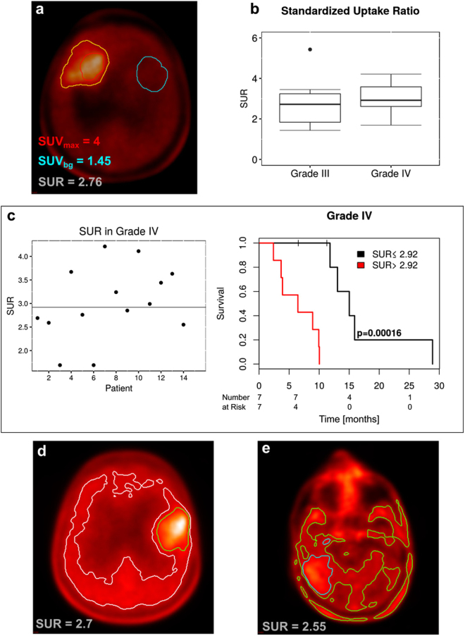
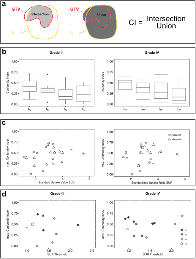
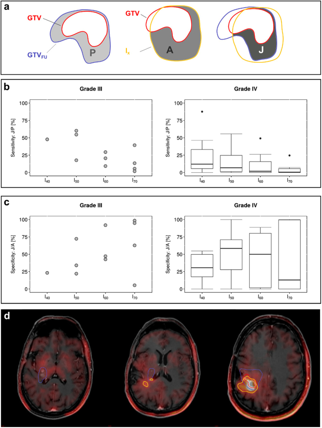
High-precision radiotherapy (HPR) of recurrent high grade glioma (HGG) requires accurate spatial allocation of these infiltrative tumors. We investigated the impact of F-FET PET on tumor delineation and progression of recurrent HGG after HPR with carbon ions. T contrast enhanced MRI and F-FET-PET scans of 26 HGG patients were fused with radiotherapy planning volumes. PET-positive (PET+) tumor volumes using different isocontours (I%) were systematically investigated and compared with MRI-derived gross tumor volumes (GTV). Standardized uptake ratios (SUR) were further correlated with GTV and tumor progression patterns. In grade IV glioma, SUR > 2.92 significantly correlated with poor median overall survival (6.5 vs 13.1 months, p = 0.00016). We found no reliable SUR cut-off criteria for definition of PET+ volumes. Overall conformity between PET and MRI-based contours was low, with maximum conformities between 0.42-0.51 at I40%. The maximum sensitivity and specificity for PET+ volumes outside of GTV predicting tumor progression were 0.16 (I40%) and 0.52 (I50%), respectively. In 75% of cases, FLAIR hyperintense area covered over 80% of PET+ volumes. F-FET-PET derived SUR has a prognostic impact in grade IV glioma. The value of substantial mismatches between MRI-based GTV and PET+ volumes to improve tumor delineation in radiotherapy awaits further validation in randomized prospective trials.

摘要

复发性高级别胶质瘤(HGG)的高精度放疗(HPR)需要准确分配这些浸润性肿瘤的空间位置。我们研究了 F-FET PET 对碳离子 HPR 后复发性 HGG 肿瘤勾画和进展的影响。26 例 HGG 患者的 T 对比增强 MRI 和 F-FET-PET 扫描与放射治疗计划体积融合。系统研究并比较了不同等剂量线(I%)的 PET 阳性(PET+)肿瘤体积与 MRI 衍生的大体肿瘤体积(GTV)。进一步将标准化摄取值(SUR)与 GTV 和肿瘤进展模式相关联。在 IV 级胶质瘤中,SUR>2.92 与较差的中位总生存期显著相关(6.5 与 13.1 个月,p=0.00016)。我们没有发现可靠的 SUR 截断标准来定义 PET+体积。PET 和基于 MRI 的轮廓之间的总体一致性较低,在 I40%时最大一致性为 0.42-0.51。预测肿瘤进展时,GTV 外 PET+体积的最大灵敏度和特异性分别为 0.16(I40%)和 0.52(I50%)。在 75%的情况下,FLAIR 高信号区覆盖了超过 80%的 PET+体积。在 IV 级胶质瘤中,F-FET-PET 衍生的 SUR 具有预后影响。在随机前瞻性试验中进一步验证之前,MRI 基于 GTV 和 PET+体积之间大量不匹配以改善放疗肿瘤勾画的价值尚待验证。

https://cdn.ncbi.nlm.nih.gov/pmc/blobs/1466/5940831/2af92e9a3d47/41598_2018_25350_Fig5_HTML.jpg
https://cdn.ncbi.nlm.nih.gov/pmc/blobs/1466/5940831/051052b87159/41598_2018_25350_Fig1_HTML.jpg
https://cdn.ncbi.nlm.nih.gov/pmc/blobs/1466/5940831/bc08f2ea53f0/41598_2018_25350_Fig2_HTML.jpg
https://cdn.ncbi.nlm.nih.gov/pmc/blobs/1466/5940831/a36ee1eea765/41598_2018_25350_Fig3_HTML.jpg
https://cdn.ncbi.nlm.nih.gov/pmc/blobs/1466/5940831/aab63f6c6b85/41598_2018_25350_Fig4_HTML.jpg
https://cdn.ncbi.nlm.nih.gov/pmc/blobs/1466/5940831/2af92e9a3d47/41598_2018_25350_Fig5_HTML.jpg

相似文献

1
Impact of F-FET PET on Target Volume Definition and Tumor Progression of Recurrent High Grade Glioma Treated with Carbon-Ion Radiotherapy.碳离子放疗治疗复发性高级别胶质瘤中 F-FET PET 对靶区定义和肿瘤进展的影响。
Sci Rep. 2018 May 8;8(1):7201. doi: 10.1038/s41598-018-25350-7.
2
Prognostic Value of O-(2-[18F]-Fluoroethyl)-L-Tyrosine-Positron Emission Tomography Imaging for Histopathologic Characteristics and Progression-Free Survival in Patients with Low-Grade Glioma.O-(2-[18F]-氟乙基)-L-酪氨酸-正电子发射断层扫描成像对低级别胶质瘤患者组织病理学特征及无进展生存期的预后价值
World Neurosurg. 2016 May;89:230-9. doi: 10.1016/j.wneu.2016.01.085. Epub 2016 Mar 9.
3
Feasibility and robustness of dynamic F-FET PET based tracer kinetic models applied to patients with recurrent high-grade glioma prior to carbon ion irradiation.适形动态 F-FET PET 示踪剂动力学模型在碳离子放疗前评估复发性高级别胶质瘤患者中的可行性和稳健性。
Sci Rep. 2018 Oct 3;8(1):14760. doi: 10.1038/s41598-018-33034-5.
4
High-grade glioma radiation therapy target volumes and patterns of failure obtained from magnetic resonance imaging and 18F-FDOPA positron emission tomography delineations from multiple observers.从多位观察者的磁共振成像和 18F-FDOPA 正电子发射断层扫描勾画中获得高级别胶质瘤放射治疗靶区和失败模式。
Int J Radiat Oncol Biol Phys. 2013 Dec 1;87(5):1100-6. doi: 10.1016/j.ijrobp.2013.09.008. Epub 2013 Oct 22.
5
Analysis of FET-PET imaging for target volume definition in patients with gliomas treated with conformal radiotherapy.分析适形放疗治疗脑胶质瘤患者时 FET-PET 成像在靶区勾画中的作用。
Radiother Oncol. 2013 Dec;109(3):487-92. doi: 10.1016/j.radonc.2013.06.043. Epub 2013 Aug 13.
6
Radiotherapy target volume definition in newly diagnosed high grade glioma using F-FET PET imaging and multiparametric perfusion MRI: A prospective study (IMAGG).使用 F-FET PET 成像和多参数灌注 MRI 对新诊断的高级别胶质瘤进行放射治疗靶区定义:一项前瞻性研究(IMAGG)。
Radiother Oncol. 2020 Sep;150:164-171. doi: 10.1016/j.radonc.2020.06.025. Epub 2020 Jun 21.
7
Comparison of F-GE-180 and dynamic F-FET PET in high grade glioma: a double-tracer pilot study.F-GE-180 与动态 F-FET PET 对比在高级别胶质瘤中的应用:一项双示踪剂初步研究。
Eur J Nucl Med Mol Imaging. 2019 Mar;46(3):580-590. doi: 10.1007/s00259-018-4166-1. Epub 2018 Sep 22.
8
An interindividual comparison of O-(2-[18F]fluoroethyl)-L-tyrosine (FET)- and L-[methyl-11C]methionine (MET)-PET in patients with brain gliomas and metastases.个体间比较 O-(2-[18F]氟乙基)-L-酪氨酸(FET)和 L-[甲基-11C]蛋氨酸(MET)-PET 在脑胶质瘤和转移瘤患者中的应用。
Int J Radiat Oncol Biol Phys. 2011 Nov 15;81(4):1049-58. doi: 10.1016/j.ijrobp.2010.07.002. Epub 2011 May 11.
9
Report of first recurrent glioma patients examined with PET-MRI prior to re-irradiation.首次复发脑胶质瘤患者再放疗前行 PET-MRI 检查报告。
PLoS One. 2019 Jul 24;14(7):e0216111. doi: 10.1371/journal.pone.0216111. eCollection 2019.
10
Assessment of various strategies for 18F-FET PET-guided delineation of target volumes in high-grade glioma patients.评估18F-FET PET引导下高级别胶质瘤患者靶区勾画的各种策略。
Eur J Nucl Med Mol Imaging. 2009 Feb;36(2):182-93. doi: 10.1007/s00259-008-0943-6. Epub 2008 Sep 26.

引用本文的文献

1
Comparison between [Ga]Ga-PSMA-617 and [F]FET PET as Imaging Biomarkers in Adult Recurrent Glioblastoma.在成人复发性胶质母细胞瘤中,[Ga]Ga-PSMA-617 与 [F]FET PET 比较作为成像生物标志物。
Int J Mol Sci. 2023 Nov 11;24(22):16208. doi: 10.3390/ijms242216208.
2
Synthetic PET via Domain Translation of 3-D MRI.通过三维磁共振成像的域转换实现合成正电子发射断层扫描
IEEE Trans Radiat Plasma Med Sci. 2023 Apr;7(4):333-343. doi: 10.1109/trpms.2022.3223275. Epub 2022 Nov 18.
3
The impact of tumor metabolic activity assessed by F-FET amino acid PET imaging in particle radiotherapy of high-grade glioma patients.

本文引用的文献

1
Towards standardization of F-FET PET imaging: do we need a consistent method of background activity assessment?迈向F-FET PET成像的标准化:我们是否需要一种一致的背景活性评估方法?
EJNMMI Res. 2017 Dec;7(1):48. doi: 10.1186/s13550-017-0295-y. Epub 2017 May 30.
2
Amino-acid PET versus MRI guided re-irradiation in patients with recurrent glioblastoma multiforme (GLIAA) - protocol of a randomized phase II trial (NOA 10/ARO 2013-1).氨基酸正电子发射断层扫描(PET)与磁共振成像(MRI)引导下复发性多形性胶质母细胞瘤(GLIAA)患者再程放疗的比较——一项随机II期试验(NOA 10/ARO 2013-1)方案
BMC Cancer. 2016 Oct 5;16(1):769. doi: 10.1186/s12885-016-2806-z.
3
F-FET氨基酸PET成像评估的肿瘤代谢活性在高级别胶质瘤患者粒子放疗中的影响
Front Oncol. 2022 Sep 20;12:901390. doi: 10.3389/fonc.2022.901390. eCollection 2022.
4
Relapsing High-Grade Glioma from Peritumoral Zone: Critical Review of Radiotherapy Treatment Options.瘤周区域复发性高级别胶质瘤:放疗治疗方案的批判性综述
Brain Sci. 2022 Mar 22;12(4):416. doi: 10.3390/brainsci12040416.
5
Whole Blood Transcriptional Fingerprints of High-Grade Glioma and Longitudinal Tumor Evolution under Carbon Ion Radiotherapy.高级别胶质瘤的全血转录指纹图谱及碳离子放疗下的肿瘤纵向演变
Cancers (Basel). 2022 Jan 28;14(3):684. doi: 10.3390/cancers14030684.
6
F-FET-PET-guided gross total resection improves overall survival in patients with WHO grade III/IV glioma: moving towards a multimodal imaging-guided resection.F-FET-PET 引导下的大体全切除可改善 WHO 分级 III/IV 级胶质瘤患者的总生存期:迈向多模态影像引导下的切除。
J Neurooncol. 2021 Oct;155(1):71-80. doi: 10.1007/s11060-021-03844-1. Epub 2021 Oct 1.
7
Potential role of functional imaging in predicting outcome for patients treated with carbon ion therapy: a review.功能成像在预测碳离子治疗患者预后中的潜在作用:综述。
Br J Radiol. 2021 Dec;94(1128):20210524. doi: 10.1259/bjr.20210524. Epub 2021 Sep 14.
8
Minicircles for a two-step blood biomarker and PET imaging early cancer detection strategy.用于两步式血液生物标志物和 PET 成像早期癌症检测策略的微小环。
J Control Release. 2021 Jul 10;335:281-289. doi: 10.1016/j.jconrel.2021.05.026. Epub 2021 May 21.
9
Charged Particle and Conventional Radiotherapy: Current Implications as Partner for Immunotherapy.带电粒子与传统放射治疗:当前作为免疫治疗搭档的意义
Cancers (Basel). 2021 Mar 23;13(6):1468. doi: 10.3390/cancers13061468.
10
Prognostic Value of microRNA-221/2 and 17-92 Families in Primary Glioblastoma Patients Treated with Postoperative Radiotherapy.原发性胶质母细胞瘤患者术后接受放疗后,miRNA-221/2 和 17-92 家族的预后价值。
Int J Mol Sci. 2021 Mar 15;22(6):2960. doi: 10.3390/ijms22062960.
Prognostic value of F-FET PET imaging in re-irradiation of high-grade glioma: Results of a phase I clinical trial.
F-FET PET成像在高级别胶质瘤再照射中的预后价值:一项I期临床试验的结果
Radiother Oncol. 2016 Oct;121(1):132-137. doi: 10.1016/j.radonc.2016.08.014. Epub 2016 Sep 9.
4
LOC283731 promoter hypermethylation prognosticates survival after radiochemotherapy in IDH1 wild-type glioblastoma patients.LOC283731启动子高甲基化可预测IDH1野生型胶质母细胞瘤患者放化疗后的生存情况。
Int J Cancer. 2016 Jul 15;139(2):424-32. doi: 10.1002/ijc.30069. Epub 2016 Mar 18.
5
ESTRO-ACROP guideline "target delineation of glioblastomas".欧洲放射肿瘤学会-头颈部肿瘤放疗协作组“胶质母细胞瘤靶区勾画”指南
Radiother Oncol. 2016 Jan;118(1):35-42. doi: 10.1016/j.radonc.2015.12.003. Epub 2016 Jan 6.
6
Volumetric Analysis of F-18-FET-PET Imaging for Brain Metastases.用于脑转移瘤的F-18-FET-PET成像的容积分析
World Neurosurg. 2015 Dec;84(6):1790-7. doi: 10.1016/j.wneu.2015.07.067. Epub 2015 Aug 6.
7
Molecular profiling of long-term survivors identifies a subgroup of glioblastoma characterized by chromosome 19/20 co-gain.对长期幸存者进行分子谱分析,确定了胶质母细胞瘤的一个亚组,其特征是染色体 19/20 共同增益。
Acta Neuropathol. 2015 Sep;130(3):419-34. doi: 10.1007/s00401-015-1427-y. Epub 2015 May 1.
8
Single-cell RNA-seq highlights intratumoral heterogeneity in primary glioblastoma.单细胞 RNA 测序凸显原发性脑胶质瘤肿瘤内异质性。
Science. 2014 Jun 20;344(6190):1396-401. doi: 10.1126/science.1254257. Epub 2014 Jun 12.
9
For the next trick: new discoveries in radiobiology applied to glioblastoma.接下来的亮点:放射生物学在胶质母细胞瘤治疗中的新发现。
Am Soc Clin Oncol Educ Book. 2014:e95-9. doi: 10.14694/EdBook_AM.2014.34.e95.
10
Analysis of FET-PET imaging for target volume definition in patients with gliomas treated with conformal radiotherapy.分析适形放疗治疗脑胶质瘤患者时 FET-PET 成像在靶区勾画中的作用。
Radiother Oncol. 2013 Dec;109(3):487-92. doi: 10.1016/j.radonc.2013.06.043. Epub 2013 Aug 13.