Alkatan Hind M, Al Otaibi Mohammad, Maktabi Azza M Y, Aljaedi Hamad, Elkhamary Sahar M, Al-Faky Yasser, Alsuhaibani Adel H
Department of Ophthalmology, King Saud University, Riyadh, Saudi Arabia.
Department of Pathology, King Saud University-Medical City, Riyadh, Saudi Arabia.
Saudi J Ophthalmol. 2018 Jan-Mar;32(1):45-51. doi: 10.1016/j.sjopt.2018.04.002. Epub 2018 Apr 5.
Hematic cyst is a rare orbital condition that has a wide range of clinical presentation and is characterized pathologically by lack of endothelial lining.
To correlate clinical and radiological features of hematic cysts, to tissue diagnosis, and investigate the possible etiology behind this condition, its relation to trauma and other interesting histopathological findings.
Retrospective case series at King Khaled Eye Specialist Hospital (KKESH) and King Abdulaziz University Hospital (KAUH) of all orbital lesions with tissue findings supporting the clinical and/or radiological diagnosis of hematic cyst.
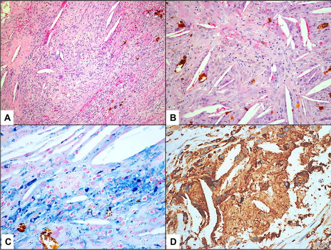
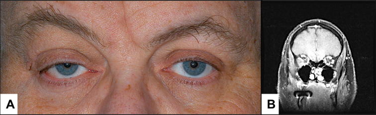
A series of 13 cases was studied, 8 males and 5 females. Age ranged from 2 to 84 years with a median of 54. Most cases presented with proptosis (76.9%) and limitation of eye movements (69.2%). History of trauma was confirmed in only 2/13. The clinical diagnosis of hematic cyst was made prior to surgery in 38.4%. Magnetic Resonance Imaging (MRI) confirmed the presence of blood in the orbit in 7/7. Surgical intervention was the mainstay of treatment. Histopathologically, these lesions demonstrated variable constituents including blood break-down products (hemosiderin), macrophages, mononuclear inflammatory cells, hemorrhage, absent endothelial lining, reactive fibrosis and capsule-like formation. Cholesterol clefts with typical granulomas and multinucleated giant cells were present in 2 cases. A clue to an underlying vascular lesion was found histopathologically in 30.8%. None of the patients developed recurrence or long-term complications with an average follow up period of 1 year.
Hematic cyst is a challenging clinical diagnosis that can be aided by radiological examination and histopathological confirmation. Trauma does not seem to play a major role while presence of a pre-existing vascular lesion with spontaneous hemorrhage may be an etiologic factor. Associated cholesterol granuloma is an interesting controversial finding. Surgical intervention is curative with possible persisting motility disturbance and/or the eye deviation and worse prognosis in post-traumatic cases.
血性囊肿是一种罕见的眼眶疾病,临床表现多样,病理特征为缺乏内皮衬里。
将血性囊肿的临床和放射学特征与组织诊断相关联,研究该疾病背后可能的病因、其与创伤的关系以及其他有趣的组织病理学发现。
在沙特国王哈立德眼科专科医院(KKESH)和阿卜杜勒阿齐兹国王大学医院(KAUH)对所有眼眶病变进行回顾性病例系列研究,这些病变的组织学检查结果支持血性囊肿的临床和/或放射学诊断。
共研究了13例病例,其中男性8例,女性5例。年龄范围为2至84岁,中位数为54岁。大多数病例表现为眼球突出(76.9%)和眼球运动受限(69.2%)。仅13例中的2例有明确的外伤史。术前临床诊断为血性囊肿的占38.4%。7例中的7例磁共振成像(MRI)证实眼眶内有血液。手术干预是主要的治疗方法。组织病理学上,这些病变表现出多种成分,包括血液分解产物(含铁血黄素)、巨噬细胞、单核炎性细胞、出血、无内皮衬里、反应性纤维化和包膜样形成。2例出现含有典型肉芽肿和多核巨细胞的胆固醇裂隙。组织病理学检查发现30.8%的病例存在潜在血管病变线索。平均随访1年,所有患者均未出现复发或长期并发症。
血性囊肿是一种具有挑战性的临床诊断,放射学检查和组织病理学确诊有助于诊断。创伤似乎不起主要作用,而存在先前的血管病变并自发性出血可能是一个病因。相关的胆固醇肉芽肿是一个有趣且有争议的发现。手术干预可治愈,但可能会持续存在眼球运动障碍和/或眼球偏斜,创伤后病例预后更差。