Samouëlian Vanessa, Mechtouf Nawel, Leblanc Eric, Cardin Guillaume B, Lhotellier Valérie, Querleu Denis, Révillion Françoise, Rodier Francis
CRCHUM et Institut du cancer de Montréal, Montreal, QC, Canada.
Université de Montréal, Département d'Obstétrique Gynécologie, Montreal, QC, Canada.
Oncotarget. 2018 Apr 24;9(31):21641-21654. doi: 10.18632/oncotarget.24956.
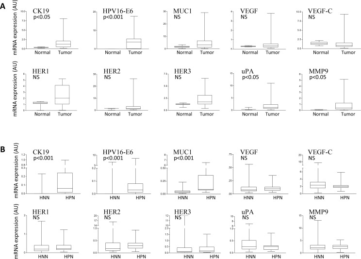
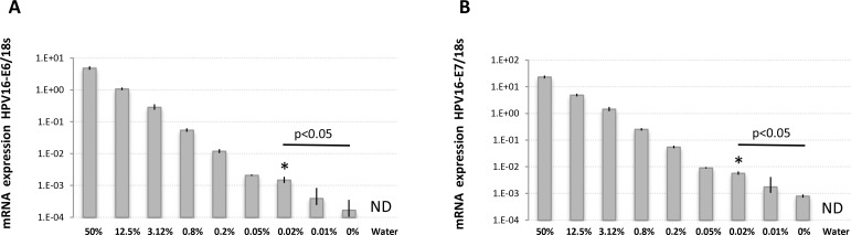
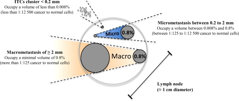
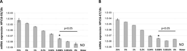
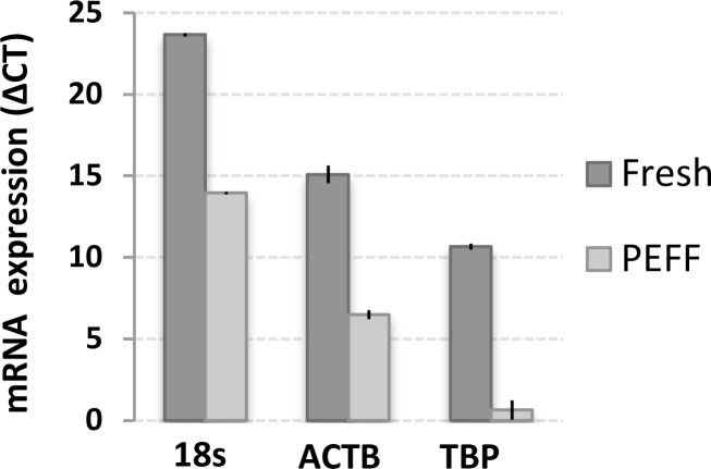
Metastatic nodal involvement is a critical prognostic factor in uterine cervical cancer (UCC). To improve current methods of detecting UCC metastases in lymph nodes (LNs), we used quantitative PCR (qPCR) to assess mRNA expression of potential metastatic biomarkers. We found that expression of HPV16-E6, cytokeratin19 (CK19), and mucin1 (MUC1) is consistently upregulated in tumors and metastatic tissues, supporting a role for these genes in UCC progression. These putative biomarkers were able to predict the presence of histologically positive metastatic LNs with respective sensitivities and specificities of 82% and 99% (CK19), 76% and 95% (HPV16-E6), and 76% and 78% (MUC1). While the biomarkers failed to detect 1.7% to 2.2% of the histologically positive LNs when used individually, combining CK19 and HPV16-E6 enhanced sensitivity and specificity to 100% and 94%, respectively. To explore the sensitivity of qPCR-based detection of varying proportions of invading HPV16-positive UCC cells, we designed a LN metastasis model that achieved a fresh cell detection limit of 0.008% (1:12500 HPV16-positive to HPV16-negative cells), and a paraffin-embedded, formalin-fixed (PEFF) detection limit of 0.02% (1:5000 HPV16-positive to HPV16-negative cells), both of which are within the theoretical detection limit for micrometastasis. Thus, HPV E6/E7 oncogenes may be useful targets for the ultrasensitive detection of nodal involvements like micrometastases in fresh or archived tissue samples. Moreover, our results suggest that the biomarker combination of CK19/HPV-E6 could support a real-time intraoperative strategy for the detection of small, but potentially lethal, metastatic nodal involvements in fresh UCC tissues.
淋巴结转移是子宫颈癌(UCC)的一个关键预后因素。为了改进当前检测UCC淋巴结(LN)转移的方法,我们使用定量聚合酶链反应(qPCR)来评估潜在转移生物标志物的mRNA表达。我们发现,HPV16-E6、细胞角蛋白19(CK19)和粘蛋白1(MUC1)在肿瘤组织和转移组织中的表达持续上调,支持这些基因在UCC进展中发挥作用。这些假定的生物标志物能够预测组织学上阳性的转移LN的存在,CK19的敏感性和特异性分别为82%和99%,HPV16-E6为76%和95%,MUC1为76%和78%。虽然这些生物标志物单独使用时未能检测出1.7%至2.2%的组织学阳性LN,但将CK19和HPV16-E6联合使用可将敏感性和特异性分别提高到100%和94%。为了探索基于qPCR检测不同比例侵袭性HPV16阳性UCC细胞的敏感性,我们设计了一个LN转移模型,新鲜细胞检测限为0.008%(HPV16阳性与HPV16阴性细胞比例为1:12500),石蜡包埋、福尔马林固定(PEFF)检测限为0.02%(HPV16阳性与HPV16阴性细胞比例为1:5000),两者均在微转移的理论检测限内。因此,HPV E6/E7癌基因可能是新鲜或存档组织样本中微转移等淋巴结受累超敏检测的有用靶点。此外,我们的结果表明,CK19/HPV-E6生物标志物组合可为新鲜UCC组织中微小但可能致命的转移淋巴结受累的实时术中检测提供支持。