Neuroscience Institute, Princeton University, Princeton, NJ 08544, USA; Electrical Engineering Department, Princeton University, Princeton, NJ 08544, USA.
Neuroscience Institute, Princeton University, Princeton, NJ 08544, USA.
Cell. 2018 May 17;173(5):1293-1306.e19. doi: 10.1016/j.cell.2018.04.040.
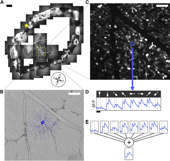
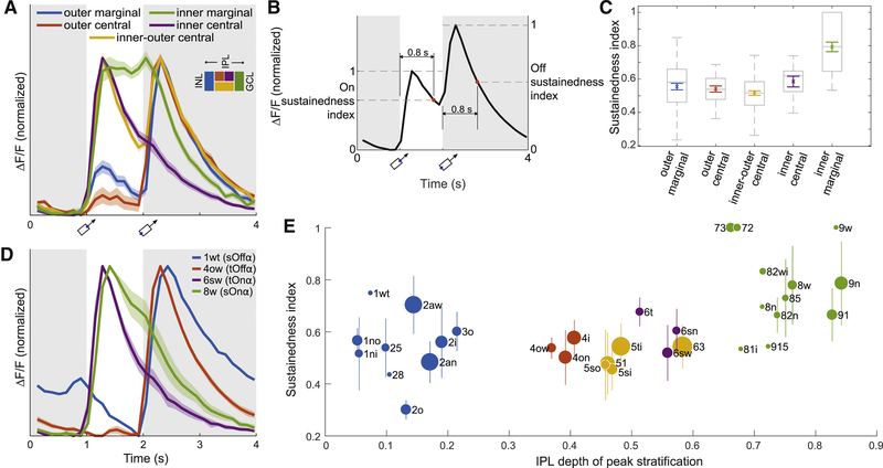
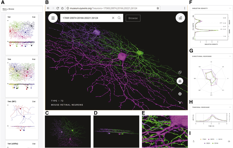
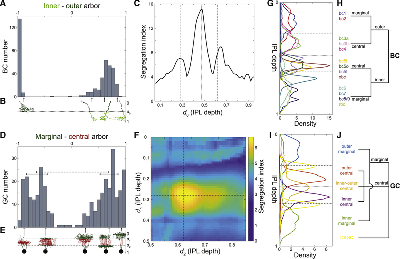
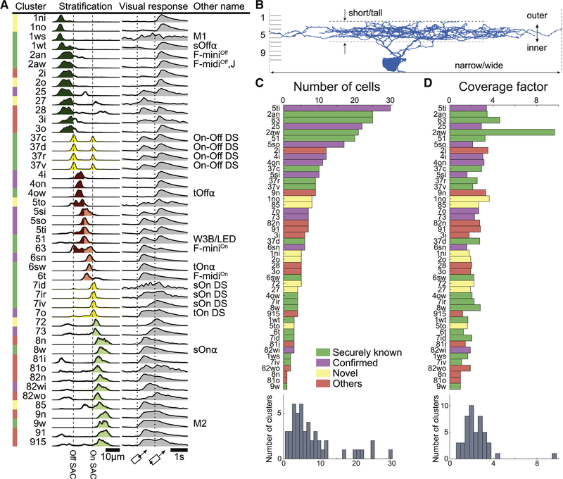
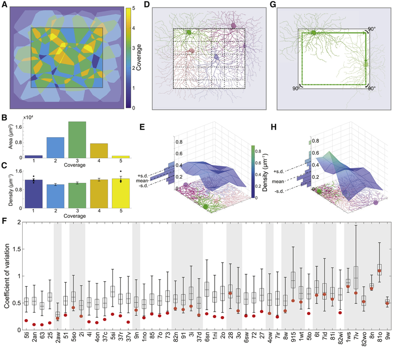
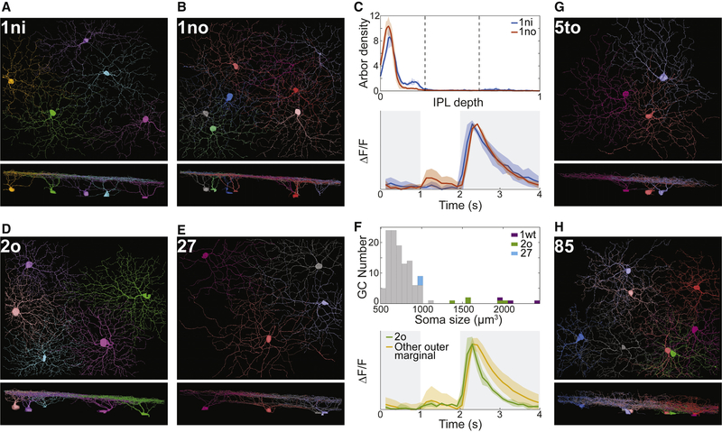
When 3D electron microscopy and calcium imaging are used to investigate the structure and function of neural circuits, the resulting datasets pose new challenges of visualization and interpretation. Here, we present a new kind of digital resource that encompasses almost 400 ganglion cells from a single patch of mouse retina. An online "museum" provides a 3D interactive view of each cell's anatomy, as well as graphs of its visual responses. The resource reveals two aspects of the retina's inner plexiform layer: an arbor segregation principle governing structure along the light axis and a density conservation principle governing structure in the tangential plane. Structure is related to visual function; ganglion cells with arbors near the layer of ganglion cell somas are more sustained in their visual responses on average. Our methods are potentially applicable to dense maps of neuronal anatomy and physiology in other parts of the nervous system.
当使用 3D 电子显微镜和钙成像技术来研究神经回路的结构和功能时,所得到的数据集提出了新的可视化和解释挑战。在这里,我们呈现了一种新的数字资源,其中包含来自单个小鼠视网膜斑块的近 400 个神经节细胞。一个在线“博物馆”提供了每个细胞解剖结构的 3D 交互式视图,以及其视觉反应的图表。该资源揭示了视网膜内丛状层的两个方面:沿光轴控制结构的分支分离原则和在切平面上控制结构的密度守恒原则。结构与视觉功能有关;树突分支靠近神经节细胞体层的神经节细胞的视觉反应平均更为持续。我们的方法可能适用于神经系统其他部位神经元解剖和生理学的密集图谱。