Division of Intramural Research, National Center for Complementary and Integrative Health, National Institutes of Health, Bethesda, MD, United States.
Faculty of Dentistry, McGill University, Montreal, QC, Canada.
Pain. 2018 Sep;159(9):1856-1866. doi: 10.1097/j.pain.0000000000001282.
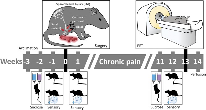
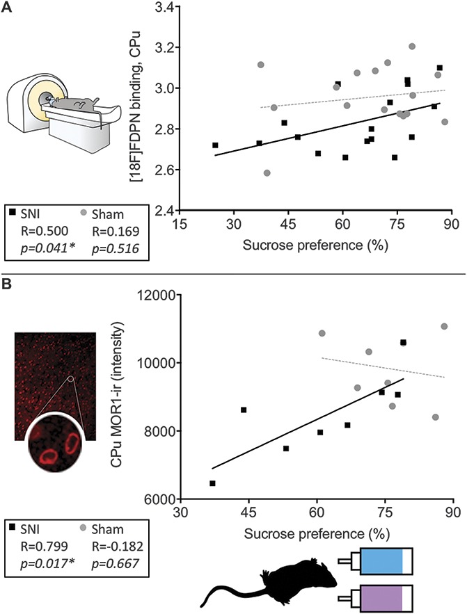
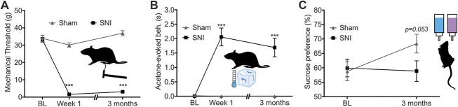
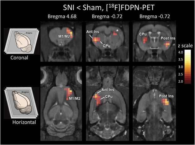
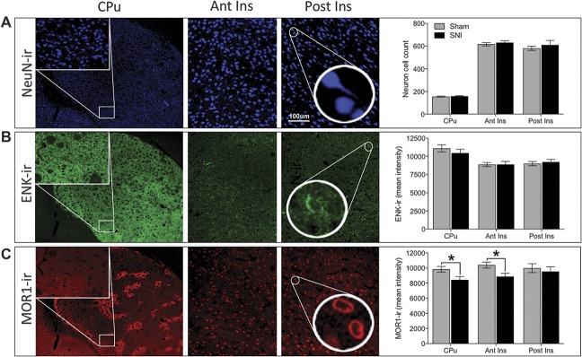
The opioid system plays a critical role in both the experience and management of pain. Although acute activation of the opioid system can lead to pain relief, the effects of chronic pain on the opioid system remain opaque. Cross-sectional positron emission tomography (PET) studies show reduced availability of brain opioid receptors in patients with chronic pain but are unable to (1) determine whether these changes are due to the chronic pain itself or due to preexisting or medication-induced differences in the endogenous opioid system, and (2) identify the neurobiological substrate of reduced opioid receptor availability. We investigated these possibilities using a well-controlled longitudinal study design in rat. Using [F]-FDPN-PET in either sham rats (n = 17) or spared nerve injury rats (n = 17), we confirmed reduced opioid receptor availability in the insula, caudate-putamen, and motor cortex of nerve injured rats 3 months after surgery, indicating that painful neuropathy altered the endogenous opioid system. Immunohistochemistry showed reduced expression of the mu-opioid receptor, MOR1, in the caudate-putamen and insula. Neither the opioid peptide enkephalin nor the neuronal marker NeuN differed between groups. In nerve-injured animals, sucrose preference, a measure of anhedonia/depression-like behavior, positively correlated with PET opioid receptor availability and MOR1-immunoreactivity in the caudate-putamen. These findings provide new evidence that the altered supraspinal opioid receptor availability observed in human patients with chronic pain may be a direct result of chronic pain. Moreover, reduced opioid receptor availability seems to reflect decreased receptor expression, which may contribute to pain-induced depression.
阿片系统在疼痛的体验和管理中起着关键作用。虽然阿片系统的急性激活可以导致疼痛缓解,但慢性疼痛对阿片系统的影响仍然不清楚。横断面正电子发射断层扫描(PET)研究表明,慢性疼痛患者大脑阿片受体的可用性降低,但无法确定这些变化是由于慢性疼痛本身还是由于内源性阿片系统的预先存在或药物诱导的差异所致,也无法确定降低阿片受体可用性的神经生物学基础。我们使用大鼠的一项精心控制的纵向研究设计来研究这些可能性。我们使用 [F]-FDPN-PET 在假手术大鼠(n = 17)或 spared 神经损伤大鼠(n = 17)中进行研究,在手术后 3 个月证实了神经损伤大鼠的岛叶、尾状核-壳核和运动皮层中的阿片受体可用性降低,表明痛性神经病改变了内源性阿片系统。免疫组织化学显示,尾状核-壳核和岛叶中的 mu-阿片受体 MOR1 的表达减少。阿片肽脑啡肽和神经元标志物 NeuN 在两组之间没有差异。在神经损伤动物中,蔗糖偏好(一种快感缺失/抑郁样行为的测量)与 PET 阿片受体可用性和尾状核-壳核中的 MOR1 免疫反应性呈正相关。这些发现提供了新的证据,表明在慢性疼痛的人类患者中观察到的改变的中枢阿片受体可用性可能是慢性疼痛的直接结果。此外,降低的阿片受体可用性似乎反映了受体表达的减少,这可能导致疼痛引起的抑郁。