Department of Ophthalmology, Northwestern University, Feinberg School of Medicine, Chicago, Illinois, United States.
Department of Ophthalmology and Optometry, Medical University of Vienna, Vienna, Austria.
Invest Ophthalmol Vis Sci. 2018 Apr 1;59(5):2167-2176. doi: 10.1167/iovs.17-23304.
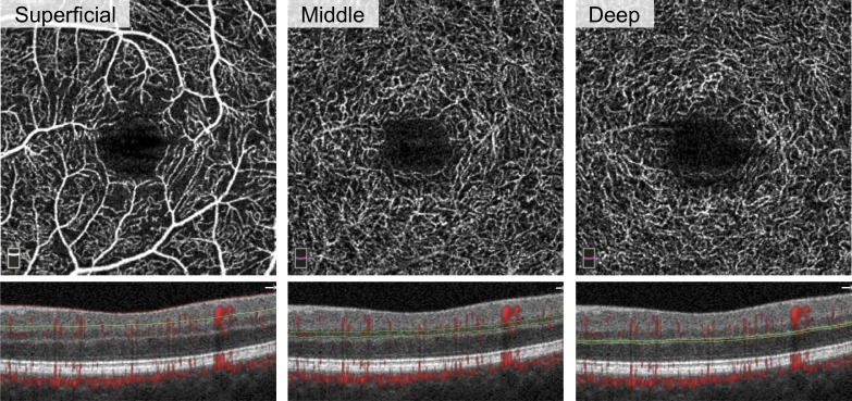
To quantify microvasculature changes in the superficial (SCP), middle (MCP), and deep capillary plexuses (DCP) in diabetic retinopathy (DR).
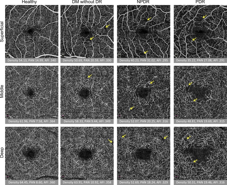
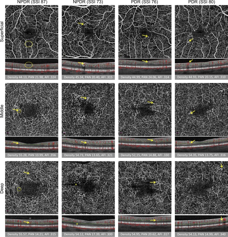
Retrospective cross-sectional study at a tertiary academic referral center, in which 26 controls (44 eyes), 27 diabetic subjects without retinopathy (44 eyes), 32 subjects with nonproliferative retinopathy (52 eyes), and 27 subjects with proliferative retinopathy (40 eyes) were imaged with optical coherence tomography angiography (OCTA). Outcome measures included parafoveal vessel density (VD), percentage area of nonperfusion (PAN), and adjusted flow index (AFI) at the different plexuses.
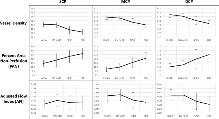
MCP VD and MCP AFI decreased with worsening DR, while PAN increased, mirroring changes within the DCP. The fitted regression line for MCP and DCP AFI were significantly different than the SCP, while DCP PAN differed from SCP PAN with disease progression. Higher SCP AFI and PAN were different in eyes with diabetes without retinopathy compared with controls. Unexpectedly, sex was found to independently influence MCP VD and AFI with worsening disease.
OCTA parameters in the MCP and DCP displayed parallel changes with DR progression, different from the SCP, emphasizing the importance of physiologic considerations in the retinal capillaries. Thus, segmentation protocols that include the MCP within the SCP may be confounded. A difference in DCP PAN with worsening DR was unmasked relative to a prior study that included the MCP with SCP. We confirm that SCP AFI and PAN may serve as early indicators of microvascular changes in DR and identify an interaction between sex and the MCP deserving further study.
定量分析糖尿病视网膜病变(DR)中浅层毛细血管丛(SCP)、中层毛细血管丛(MCP)和深层毛细血管丛(DCP)的微血管变化。
这是一项在三级学术转诊中心进行的回顾性横断面研究,纳入 26 名对照者(44 只眼)、27 名无糖尿病视网膜病变的糖尿病患者(44 只眼)、32 名非增殖性视网膜病变患者(52 只眼)和 27 名增殖性视网膜病变患者(40 只眼),所有患者均接受光学相干断层扫描血管造影(OCTA)检查。主要观察指标包括黄斑区旁中心凹血管密度(VD)、无灌注面积百分比(PAN)和不同丛的校正血流指数(AFI)。
随着 DR 病情的恶化,MCP VD 和 MCP AFI 逐渐降低,而 PAN 逐渐增加,与 DCP 内的变化相似。MCP 和 DCP 的拟合回归线与 SCP 明显不同,而 DCP PAN 随着疾病的进展与 SCP PAN 不同。与对照组相比,无糖尿病视网膜病变的糖尿病患者的 SCP AFI 和 PAN 更高。出乎意料的是,性别与疾病的恶化独立影响 MCP VD 和 AFI。
与 SCP 相比,MCP 和 DCP 的 OCTA 参数在 DR 进展过程中呈现出平行变化,强调了视网膜毛细血管的生理考虑的重要性。因此,包括 MCP 在内的 SCP 分段方案可能会受到干扰。与之前包含 SCP 和 MCP 的研究相比,DCP PAN 随着 DR 病情的恶化而出现的差异更加明显。我们证实 SCP AFI 和 PAN 可能作为 DR 微血管变化的早期指标,并发现性别与 MCP 之间的相互作用值得进一步研究。