• 文献检索
  • 文档翻译
  • 深度研究
  • 学术资讯
  • Suppr Zotero 插件Zotero 插件
  • 邀请有礼
  • 套餐&价格
  • 历史记录
应用&插件
Suppr Zotero 插件Zotero 插件浏览器插件Mac 客户端Windows 客户端微信小程序
定价
高级版会员购买积分包购买API积分包
服务
文献检索文档翻译深度研究API 文档MCP 服务
关于我们
关于 Suppr公司介绍联系我们用户协议隐私条款
关注我们

Suppr 超能文献

核心技术专利:CN118964589B侵权必究
粤ICP备2023148730 号-1Suppr @ 2026

文献检索

告别复杂PubMed语法,用中文像聊天一样搜索,搜遍4000万医学文献。AI智能推荐,让科研检索更轻松。

立即免费搜索

文件翻译

保留排版,准确专业,支持PDF/Word/PPT等文件格式,支持 12+语言互译。

免费翻译文档

深度研究

AI帮你快速写综述,25分钟生成高质量综述,智能提取关键信息,辅助科研写作。

立即免费体验

利用动态对比增强磁共振成像得出的药代动力学参数对胰腺神经内分泌肿瘤进行分级

Grading of pancreatic neuroendocrine neoplasms using pharmacokinetic parameters derived from dynamic contrast-enhanced MRI.

作者信息

Zhao Weiwei, Quan Zhiyong, Huang Xufang, Ren Jing, Wen Didi, Zhang Guangwen, Shi Zhongqiang, Yin Hong, Huan Yi

机构信息

Department of Radiology, Xijing Hospital, Fourth Military Medical University, Xi'an, Shaanxi 710032, P.R. China.

Department of Nuclear Medicine, Xijing Hospital, Fourth Military Medical University, Xi'an, Shaanxi 710032, P.R. China.

出版信息

Oncol Lett. 2018 Jun;15(6):8349-8356. doi: 10.3892/ol.2018.8384. Epub 2018 Mar 29.

DOI:10.3892/ol.2018.8384
PMID:29805568
原文链接:https://pmc.ncbi.nlm.nih.gov/articles/PMC5950181/
Abstract

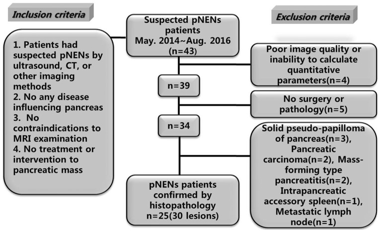
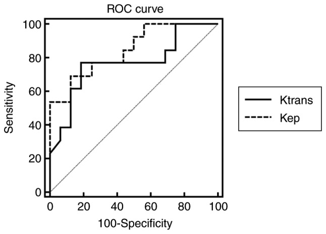
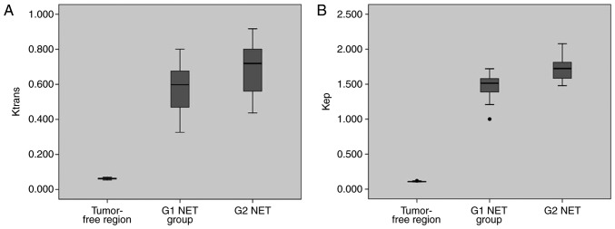
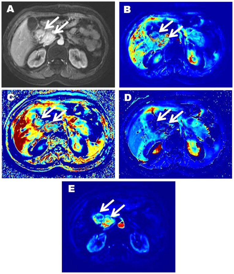
The present study aimed to evaluate the diagnostic efficacy of pharmacokinetic parameters derived from dynamic contrast-enhanced magnetic resonance imaging (DCE-MRI) in prospective evaluation of pancreatic neuroendocrine neoplasms (pNENs) grading. A total of 25 histologically proven patients with pNENs (30 lesions in total) who underwent DCE-MRI were enrolled. Lesions were divided into G1, G2 neuroendocrine tumor (NET) and G3 NET/neuroendocrine carcinoma (NEC) groups based on their histological findings according to 2017 World Health Organization Neuroendocrine Tumor Classification Guideline. In addition, the same numbers of tumor-free regions were selected using as normal control group. For each group, pharmacokinetic DCE parameters: volume transfer constant (); contrast transfer rate constant (); extravascular extracellular space volume fraction (v); and plasma volume fraction (v) were calculated with Extended Tofts Linear model. Receiver operator characteristics analysis was conducted to assess the diagnostic efficacy of these parameters in pNENs grading. There were significant differences of , , v and v between tumor-free areas and G1, G2 NET (P<0.001). The and of G1 NET were significantly lower compared with those of G2 ones (P<0.005). The area under the curve of and in differentiating G2 from G1 NET were 0.767 and 0.846, respectively. When was >0.667 and k >1.644, the sensitivity of diagnosing G2 NET was the lowest (53.85%), but the specificity was the highest (93.75%). When was >0.667 or >1.644, the sensitivity of diagnosing G2 NET was 92.31%, but the specificity was 75.00%. Pharmacokinetic parameters of DCE-MRI, particularly the quantitative values of and , are helpful for differentiating G2 NET from G1 ones.

摘要

本研究旨在评估动态对比增强磁共振成像(DCE-MRI)得出的药代动力学参数在前瞻性评估胰腺神经内分泌肿瘤(pNENs)分级中的诊断效能。共有25例经组织学证实的pNENs患者(共30个病灶)接受了DCE-MRI检查并被纳入研究。根据2017年世界卫生组织神经内分泌肿瘤分类指南,依据组织学结果将病灶分为G1、G2神经内分泌肿瘤(NET)以及G3 NET/神经内分泌癌(NEC)组。此外,选取相同数量的无肿瘤区域作为正常对照组。对于每组,采用扩展Tofts线性模型计算药代动力学DCE参数:容积转运常数();对比剂转运速率常数();血管外细胞外间隙容积分数(v);以及血浆容积分数(v)。进行受试者操作特征分析以评估这些参数在pNENs分级中的诊断效能。无肿瘤区域与G1、G2 NET之间的、、v和v存在显著差异(P<0.001)。G1 NET的和显著低于G2 NET(P<0.005)。在区分G2与G1 NET时,和的曲线下面积分别为0.767和0.846。当>0.667且k>1.644时,诊断G2 NET的敏感性最低(53.85%),但特异性最高(93.75%)。当>0.667或>1.644时,诊断G2 NET的敏感性为92.31%,但特异性为75.00%。DCE-MRI的药代动力学参数,尤其是和的定量值,有助于区分G2 NET与G1 NET。

https://cdn.ncbi.nlm.nih.gov/pmc/blobs/f4a2/5950181/7b279dd1a977/ol-15-06-8349-g04.jpg
https://cdn.ncbi.nlm.nih.gov/pmc/blobs/f4a2/5950181/4999415b5767/ol-15-06-8349-g00.jpg
https://cdn.ncbi.nlm.nih.gov/pmc/blobs/f4a2/5950181/7039696979da/ol-15-06-8349-g01.jpg
https://cdn.ncbi.nlm.nih.gov/pmc/blobs/f4a2/5950181/ba0b0e6ff758/ol-15-06-8349-g02.jpg
https://cdn.ncbi.nlm.nih.gov/pmc/blobs/f4a2/5950181/642a321b659d/ol-15-06-8349-g03.jpg
https://cdn.ncbi.nlm.nih.gov/pmc/blobs/f4a2/5950181/7b279dd1a977/ol-15-06-8349-g04.jpg

相似文献

1
Grading of pancreatic neuroendocrine neoplasms using pharmacokinetic parameters derived from dynamic contrast-enhanced MRI.利用动态对比增强磁共振成像得出的药代动力学参数对胰腺神经内分泌肿瘤进行分级
Oncol Lett. 2018 Jun;15(6):8349-8356. doi: 10.3892/ol.2018.8384. Epub 2018 Mar 29.
2
High-temporal resolution DCE-MRI improves assessment of intra- and peri-breast lesions categorized as BI-RADS 4.高时间分辨率 DCE-MRI 提高了 BI-RADS 4 类乳腺内及周围病变的评估能力。
BMC Med Imaging. 2023 Apr 19;23(1):58. doi: 10.1186/s12880-023-01015-4.
3
Quantitative analysis of permeability for glioma grading using dynamic contrast-enhanced magnetic resonance imaging.使用动态对比增强磁共振成像对胶质瘤分级的渗透性进行定量分析。
Oncol Lett. 2017 Nov;14(5):5418-5426. doi: 10.3892/ol.2017.6895. Epub 2017 Sep 6.
4
Dynamic contrast-enhanced magnetic resonance imaging and diffusion-weighted imaging in the activity staging of terminal ileum Crohn's disease.动态对比增强磁共振成像和扩散加权成像在末端回肠克罗恩病活动分期中的应用
World J Gastroenterol. 2020 Oct 21;26(39):6057-6073. doi: 10.3748/wjg.v26.i39.6057.
5
Pharmacokinetic analysis for the differentiation of pituitary microadenoma subtypes through dynamic contrast-enhanced magnetic resonance imaging.通过动态对比增强磁共振成像对垂体微腺瘤亚型进行鉴别的药代动力学分析。
Oncol Lett. 2019 May;17(5):4237-4244. doi: 10.3892/ol.2019.10083. Epub 2019 Feb 28.
6
Assessment of liver fibrosis using pharmacokinetic parameters of dynamic contrast-enhanced magnetic resonance imaging.利用动态对比增强磁共振成像的药代动力学参数评估肝纤维化
J Magn Reson Imaging. 2016 Jul;44(1):98-104. doi: 10.1002/jmri.25132. Epub 2015 Dec 28.
7
Evaluating the Relationship Between Dynamic Contrast-Enhanced MRI (DCE-MRI) Parameters and Pathological Characteristics in Breast Cancer.评估动态对比增强磁共振成像(DCE-MRI)参数与乳腺癌病理特征之间的关系。
J Magn Reson Imaging. 2020 Nov;52(5):1360-1373. doi: 10.1002/jmri.27241. Epub 2020 Jun 10.
8
Extracellular volume fraction with MRI: As an alternative predictive biomarker to dynamic contrast-enhanced MRI for chemotherapy response of pancreatic ductal adenocarcinoma.MRI 下细胞外容积分数:作为预测胰腺导管腺癌化疗反应的替代生物标志物,优于动态对比增强 MRI。
Eur J Radiol. 2021 Dec;145:110036. doi: 10.1016/j.ejrad.2021.110036. Epub 2021 Nov 16.
9
Repeatability and correlations of dynamic contrast enhanced and T2* MRI in patients with advanced pancreatic ductal adenocarcinoma.晚期胰腺导管腺癌患者动态对比增强磁共振成像与T2*磁共振成像的可重复性及相关性
Magn Reson Imaging. 2018 Jul;50:1-9. doi: 10.1016/j.mri.2018.02.005. Epub 2018 Feb 21.
10
Quantitative dynamic contrast-enhanced magnetic resonance imaging in a VX2 rabbit liver tumour model using different gadolinium-based contrast agents: comparison of DCE-MRI quantitative results between Magnevist and Eovist.使用不同钆基造影剂的VX2兔肝肿瘤模型中的定量动态对比增强磁共振成像:马根维显和欧乃影的DCE-MRI定量结果比较
BJR Case Rep. 2017 Apr 12;3(3):20160099. doi: 10.1259/bjrcr.20160099. eCollection 2017.

引用本文的文献

1
The multidisciplinary team for gastroenteropancreatic neuroendocrine tumours: the radiologist's challenge.胃肠胰神经内分泌肿瘤的多学科团队:放射科医生的挑战。
Radiol Oncol. 2019 Oct 25;53(4):373-387. doi: 10.2478/raon-2019-0040.
2
CT radiomics may predict the grade of pancreatic neuroendocrine tumors: a multicenter study.CT 放射组学可预测胰腺神经内分泌肿瘤的分级:一项多中心研究。
Eur Radiol. 2019 Dec;29(12):6880-6890. doi: 10.1007/s00330-019-06176-x. Epub 2019 Jun 21.
3
Grading of Neuroendocrine Carcinomas: Correlation of Ga-PET/CT Scan with Tissue Biomarkers.

本文引用的文献

1
Recent updates on grading and classification of neuroendocrine tumors.神经内分泌肿瘤分级与分类的最新进展
Ann Diagn Pathol. 2017 Aug;29:11-16. doi: 10.1016/j.anndiagpath.2017.04.005. Epub 2017 Apr 13.
2
Management of neuroendocrine carcinomas of the pancreas (WHO G3): A tailored approach between proliferation and morphology.胰腺神经内分泌癌(世界卫生组织G3)的管理:增殖与形态之间的个体化方法。
World J Gastroenterol. 2016 Dec 7;22(45):9944-9953. doi: 10.3748/wjg.v22.i45.9944.
3
Long-term outcomes and prognostic factors in neuroendocrine carcinomas of the pancreas: Morphology matters.
神经内分泌癌分级:Ga-PET/CT 扫描与组织生物标志物的相关性。
Dis Markers. 2018 Dec 2;2018:6878409. doi: 10.1155/2018/6878409. eCollection 2018.
胰腺神经内分泌癌的长期预后及预后因素:形态学至关重要。
Surgery. 2016 Mar;159(3):862-71. doi: 10.1016/j.surg.2015.09.012.
4
Optimal cut-off value of perfusion parameters for diagnosing prostate cancer and for assessing aggressiveness associated with Gleason score.用于诊断前列腺癌以及评估与 Gleason 评分相关的侵袭性的灌注参数的最佳截断值。
Clin Imaging. 2015 Sep-Oct;39(5):834-40. doi: 10.1016/j.clinimag.2015.04.020. Epub 2015 May 7.
5
Dynamic Contrast-Enhanced Perfusion MRI of High Grade Brain Gliomas Obtained with Arterial or Venous Waveform Input Function.采用动脉或静脉波形输入函数获得的高级别脑胶质瘤动态对比增强灌注磁共振成像
J Neuroimaging. 2016 Jan-Feb;26(1):124-9. doi: 10.1111/jon.12254. Epub 2015 Apr 29.
6
Dynamic contrast enhanced MRI-derived parameters are potential biomarkers of therapeutic response in bladder carcinoma.动态对比增强磁共振成像衍生参数是膀胱癌治疗反应的潜在生物标志物。
Eur J Radiol. 2015 Jun;84(6):1023-8. doi: 10.1016/j.ejrad.2015.02.026. Epub 2015 Mar 6.
7
Neuroendocrine neoplasms of the pancreas at dynamic enhanced CT: comparison between grade 3 neuroendocrine carcinoma and grade 1/2 neuroendocrine tumour.胰腺神经内分泌肿瘤的动态增强 CT 表现:3 级神经内分泌癌与 1/2 级神经内分泌肿瘤的比较。
Eur Radiol. 2015 May;25(5):1375-83. doi: 10.1007/s00330-014-3532-z. Epub 2014 Dec 3.
8
Intermodel agreement of myocardial blood flow estimation from stress-rest myocardial perfusion magnetic resonance imaging in patients with coronary artery disease.冠心病患者应力-静息心肌灌注磁共振成像估测心肌血流的不同模式间一致性
Invest Radiol. 2015 Apr;50(4):275-82. doi: 10.1097/RLI.0000000000000114.
9
Dynamic contrast-enhanced MRI of gastric cancer: Correlation of the perfusion parameters with pathological prognostic factors.胃癌的动态对比增强磁共振成像:灌注参数与病理预后因素的相关性。
J Magn Reson Imaging. 2015 Jun;41(6):1608-14. doi: 10.1002/jmri.24711. Epub 2014 Jul 16.
10
Assessment of early response to concurrent chemoradiotherapy in cervical cancer: value of diffusion-weighted and dynamic contrast-enhanced MR imaging.宫颈癌同步放化疗早期反应的评估:扩散加权成像与动态对比增强磁共振成像的价值
Magn Reson Imaging. 2014 Oct;32(8):993-1000. doi: 10.1016/j.mri.2014.05.009. Epub 2014 Jun 23.