• 文献检索
  • 文档翻译
  • 深度研究
  • 学术资讯
  • Suppr Zotero 插件Zotero 插件
  • 邀请有礼
  • 套餐&价格
  • 历史记录
应用&插件
Suppr Zotero 插件Zotero 插件浏览器插件Mac 客户端Windows 客户端微信小程序
定价
高级版会员购买积分包购买API积分包
服务
文献检索文档翻译深度研究API 文档MCP 服务
关于我们
关于 Suppr公司介绍联系我们用户协议隐私条款
关注我们

Suppr 超能文献

核心技术专利:CN118964589B侵权必究
粤ICP备2023148730 号-1Suppr @ 2026

文献检索

告别复杂PubMed语法,用中文像聊天一样搜索,搜遍4000万医学文献。AI智能推荐,让科研检索更轻松。

立即免费搜索

文件翻译

保留排版,准确专业,支持PDF/Word/PPT等文件格式,支持 12+语言互译。

免费翻译文档

深度研究

AI帮你快速写综述,25分钟生成高质量综述,智能提取关键信息,辅助科研写作。

立即免费体验

使用动态对比增强磁共振成像对胶质瘤分级的渗透性进行定量分析。

Quantitative analysis of permeability for glioma grading using dynamic contrast-enhanced magnetic resonance imaging.

作者信息

Zhao Ming, Guo Li-Li, Huang Ning, Wu Qiong, Zhou Li, Zhao Hui, Zhang Jing, Fu Kuang

机构信息

Department of MR Diagnosis, The Second Affiliated Hospital of Harbin Medical University, Harbin, Heilongjiang 150086, P.R. China.

Life Science, GE Healthcare Life Sciences China, Beijing 100176, P.R. China.

出版信息

Oncol Lett. 2017 Nov;14(5):5418-5426. doi: 10.3892/ol.2017.6895. Epub 2017 Sep 6.

DOI:10.3892/ol.2017.6895
PMID:29113174
原文链接:https://pmc.ncbi.nlm.nih.gov/articles/PMC5656018/
Abstract

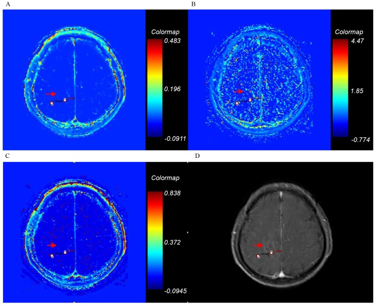
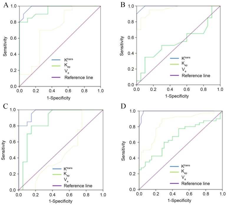
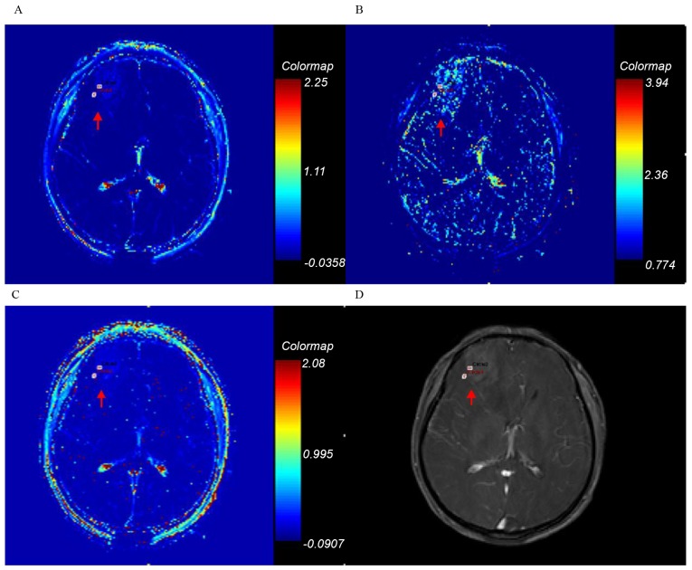
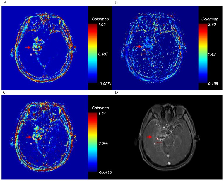
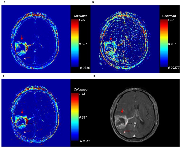
The objective of the present study was to quantitatively analyze the permeability of tumor entity and peritumor edema in glioma grading, using dynamic contrast-enhanced magnetic resonance imaging (DCE-MRI). In the present retrospective study, 80 patients underwent T1-weighted DCE-MRI examination at 3.0 T and the pathological results (including astrocytoma and oligodendroglioma) were obtained between January 2012 and June 2015. All cases were surgically validated as grade I-IV gliomas. The original DCE-MRI data were analyzed using dual compartment modified Tofts model. The forward volume transfer constant (K), backflux rate (k) and fractional volume (v) were calculated with the region of interest selected on the highest permeability area of the tumor entity and peritumor edema. Analysis of variance with the Bonferroni correction was used to compare the values of K, k, and v of the tumor entity and peritumor edema in different glioma grades. The results of the present study revealed that the K, k, and v values in each stage were associated with the pathological grading (r=0.951, 0.804 and 0.766, respectively). There were significant differences identified between different tumor grades in K, k, with the exception being between grades II and III in k. In addition, there was a significant difference revealed between grade I/II and grade III/IV in v. Receiver operator characteristics curve analysis was used to evaluate the diagnosis accuracies of permeability parameters. K was demonstrated to exhibit the highest sensitivity and specificity for evaluating the tumor grade. With the threshold values of 0.160, 0.420 and 0.935 in K on tumor, glioma grades I vs. II, II vs III and III vs. IV may be differentiated with sensitivities of 0.900, 0.950 and 0.950, and specificities of 0.950, 0.950 and 0.850, respectively. Furthermore, associations were observed between the K, k and v values of peritumor edema and the pathological grading in glioma (K r=0.438, P<0.001; K r=0.385, P<0.001; V r=0.397, P<0.001, respectively). K values in peritumoral edema revealed significant differences between low-grade and high-grade glioma. The sensitivity and specificity for K of peritumor edema were 0.975 and 0.950, with a threshold value of 0.007. Therefore, the DCE-MRI parameters of K of tumor entity and peritumor edema in gliomas may be used to accurately differentiate glioma grades.

摘要

本研究的目的是使用动态对比增强磁共振成像(DCE-MRI)对神经胶质瘤分级中肿瘤实体和瘤周水肿的通透性进行定量分析。在本回顾性研究中,80例患者于2012年1月至2015年6月期间接受了3.0 T的T1加权DCE-MRI检查,并获得了病理结果(包括星形细胞瘤和少突胶质细胞瘤)。所有病例均经手术验证为I-IV级神经胶质瘤。使用双室改良Tofts模型分析原始DCE-MRI数据。在前瞻性容积转移常数(K)、回流率(k)和容积分数(v)的计算中,感兴趣区域选在肿瘤实体和瘤周水肿的最高通透性区域。采用经Bonferroni校正的方差分析来比较不同神经胶质瘤分级中肿瘤实体和瘤周水肿的K、k和v值。本研究结果显示,各阶段的K、k和v值与病理分级相关(r分别为0.951、0.804和0.766)。不同肿瘤分级之间的K、k存在显著差异,k在II级和III级之间除外。此外,I/II级和III/IV级之间的v存在显著差异。采用受试者工作特征曲线分析来评估通透性参数的诊断准确性。结果表明,K对评估肿瘤分级具有最高的敏感性和特异性。以肿瘤K值的阈值0.160、0.420和0.935,可分别区分I级与II级、II级与III级、III级与IV级神经胶质瘤,敏感性分别为0.900、0.950和0.950,特异性分别为0.950、0.950和0.850。此外,观察到瘤周水肿的K、k和v值与神经胶质瘤的病理分级之间存在相关性(K r = 0.438,P < 0.001;k r = 0.385,P < 0.001;v r = 0.397,P < 0.001)。瘤周水肿的K值在低级别和高级别神经胶质瘤之间存在显著差异。瘤周水肿K值的敏感性和特异性分别为0.975和0.950,阈值为0.007。因此,神经胶质瘤中肿瘤实体和瘤周水肿的DCE-MRI参数K可用于准确区分神经胶质瘤分级。

https://cdn.ncbi.nlm.nih.gov/pmc/blobs/7c6c/5656018/aa048c8e9ed2/ol-14-05-5418-g04.jpg
https://cdn.ncbi.nlm.nih.gov/pmc/blobs/7c6c/5656018/2d28cbb7432a/ol-14-05-5418-g00.jpg
https://cdn.ncbi.nlm.nih.gov/pmc/blobs/7c6c/5656018/cd95edc7a9ce/ol-14-05-5418-g01.jpg
https://cdn.ncbi.nlm.nih.gov/pmc/blobs/7c6c/5656018/88cdfd7df89a/ol-14-05-5418-g02.jpg
https://cdn.ncbi.nlm.nih.gov/pmc/blobs/7c6c/5656018/f16df3644df5/ol-14-05-5418-g03.jpg
https://cdn.ncbi.nlm.nih.gov/pmc/blobs/7c6c/5656018/aa048c8e9ed2/ol-14-05-5418-g04.jpg

相似文献

1
Quantitative analysis of permeability for glioma grading using dynamic contrast-enhanced magnetic resonance imaging.使用动态对比增强磁共振成像对胶质瘤分级的渗透性进行定量分析。
Oncol Lett. 2017 Nov;14(5):5418-5426. doi: 10.3892/ol.2017.6895. Epub 2017 Sep 6.
2
[Diagnostic Value of Combining Permeability with T1 Perfusion Parameters in Quantitative Dynamic Contrast-enhanced Magnetic Resonance Imaging for Glioma Grading].[渗透性与T1灌注参数相结合在胶质瘤分级定量动态对比增强磁共振成像中的诊断价值]
Zhongguo Yi Xue Ke Xue Yuan Xue Bao. 2015 Dec;37(6):674-80. doi: 10.3881/j.issn.1000-503X.2015.06.007.
3
Diagnostic Values of DCE-MRI and DSC-MRI for Differentiation Between High-grade and Low-grade Gliomas: A Comprehensive Meta-analysis.DCE-MRI 和 DSC-MRI 对高级别和低级别胶质瘤鉴别诊断的价值:一项综合荟萃分析。
Acad Radiol. 2018 Mar;25(3):338-348. doi: 10.1016/j.acra.2017.10.001. Epub 2017 Dec 6.
4
Dynamic contrast-enhanced magnetic resonance imaging and diffusion-weighted imaging in the activity staging of terminal ileum Crohn's disease.动态对比增强磁共振成像和扩散加权成像在末端回肠克罗恩病活动分期中的应用
World J Gastroenterol. 2020 Oct 21;26(39):6057-6073. doi: 10.3748/wjg.v26.i39.6057.
5
Glioma grading capability: comparisons among parameters from dynamic contrast-enhanced MRI and ADC value on DWI.基于动态对比增强 MRI 参数和 DWI 上 ADC 值的胶质瘤分级能力比较。
Korean J Radiol. 2013 May-Jun;14(3):487-92. doi: 10.3348/kjr.2013.14.3.487. Epub 2013 May 2.
6
Effects of artery input function on dynamic contrast-enhanced MRI for determining grades of gliomas.动脉内输入函数对动态对比增强磁共振成像确定胶质瘤分级的影响。
Br J Radiol. 2021 Mar 1;94(1119):20200699. doi: 10.1259/bjr.20200699. Epub 2020 Dec 17.
7
Microvascular permeability of brain astrocytoma with contrast-enhanced magnetic resonance imaging: correlation analysis with histopathologic grade.脑星形细胞瘤的磁共振对比增强成像的微血管通透性:与组织病理学分级的相关性分析。
Chin Med J (Engl). 2013;126(10):1953-6.
8
Evaluating the Relationship Between Dynamic Contrast-Enhanced MRI (DCE-MRI) Parameters and Pathological Characteristics in Breast Cancer.评估动态对比增强磁共振成像(DCE-MRI)参数与乳腺癌病理特征之间的关系。
J Magn Reson Imaging. 2020 Nov;52(5):1360-1373. doi: 10.1002/jmri.27241. Epub 2020 Jun 10.
9
Prognostic value of preoperative dynamic contrast-enhanced MRI perfusion parameters for high-grade glioma patients.术前动态对比增强磁共振成像灌注参数对高级别胶质瘤患者的预后价值
Neuroradiology. 2016 Dec;58(12):1197-1208. doi: 10.1007/s00234-016-1741-7. Epub 2016 Oct 29.
10
Dynamic contrast-enhanced and dynamic susceptibility contrast perfusion MR imaging for glioma grading: Preliminary comparison of vessel compartment and permeability parameters using hotspot and histogram analysis.动态对比增强和动态磁敏感对比灌注磁共振成像用于胶质瘤分级:使用热点和直方图分析对血管腔室和通透性参数的初步比较
Eur J Radiol. 2016 Jun;85(6):1147-56. doi: 10.1016/j.ejrad.2016.03.020. Epub 2016 Mar 22.

引用本文的文献

1
A multiparametric perspective on C6 and F98 cell lines in orthotopic rat models for glioblastoma research.胶质母细胞瘤研究原位大鼠模型中C6和F98细胞系的多参数视角
Sci Rep. 2025 Jul 2;15(1):22547. doi: 10.1038/s41598-025-06684-5.
2
Review of tracer kinetic models in evaluation of gliomas using dynamic contrast-enhanced imaging.使用动态对比增强成像评估神经胶质瘤的示踪剂动力学模型综述。
Front Oncol. 2024 Jun 14;14:1380793. doi: 10.3389/fonc.2024.1380793. eCollection 2024.
3
Immune cell identity behind the K mapping of mouse glioblastoma.

本文引用的文献

1
Dynamic Contrast-Enhanced Perfusion MRI of High Grade Brain Gliomas Obtained with Arterial or Venous Waveform Input Function.采用动脉或静脉波形输入函数获得的高级别脑胶质瘤动态对比增强灌注磁共振成像
J Neuroimaging. 2016 Jan-Feb;26(1):124-9. doi: 10.1111/jon.12254. Epub 2015 Apr 29.
2
Discrimination between glioma grades II and III in suspected low-grade gliomas using dynamic contrast-enhanced and dynamic susceptibility contrast perfusion MR imaging: a histogram analysis approach.利用动态对比增强和动态磁敏感对比灌注磁共振成像,通过直方图分析方法鉴别疑似低级别胶质瘤中的二级和三级胶质瘤。
Neuroradiology. 2014 Dec;56(12):1031-8. doi: 10.1007/s00234-014-1426-z. Epub 2014 Sep 10.
3
小鼠神经胶质瘤 K 映射背后的免疫细胞特征。
Magn Reson Imaging. 2023 Nov;103:92-101. doi: 10.1016/j.mri.2023.06.008. Epub 2023 Jun 21.
4
Imaging biomarkers for clinical applications in neuro-oncology: current status and future perspectives.神经肿瘤学临床应用中的影像学生物标志物:现状与未来展望。
Biomark Res. 2023 Mar 29;11(1):35. doi: 10.1186/s40364-023-00476-7.
5
Peritumor Edema Serves as an Independent Predictive Factor of Recurrence Patterns and Recurrence-Free Survival for High-Grade Glioma.瘤周水肿可作为高级别胶质瘤复发模式和无复发生存的独立预测因素。
Comput Math Methods Med. 2022 Jul 27;2022:9547166. doi: 10.1155/2022/9547166. eCollection 2022.
6
The Value of Dynamic Contrast-Enhanced Magnetic Resonance Imaging (DCE-MRI) in the Differentiation of Pseudoprogression and Recurrence of Intracranial Gliomas.动态对比增强磁共振成像(DCE-MRI)在颅内胶质瘤假性进展和复发鉴别中的价值。
Contrast Media Mol Imaging. 2022 Jul 22;2022:5680522. doi: 10.1155/2022/5680522. eCollection 2022.
7
Standard clinical approaches and emerging modalities for glioblastoma imaging.胶质母细胞瘤成像的标准临床方法和新兴模式
Neurooncol Adv. 2022 May 26;4(1):vdac080. doi: 10.1093/noajnl/vdac080. eCollection 2022 Jan-Dec.
8
Nomograms for predicting the overall survival of patients with cerebellar glioma: an analysis of the surveillance epidemiology and end results (SEER) database.预测小脑胶质瘤患者总生存期的列线图:监测、流行病学和最终结果(SEER)数据库分析。
Sci Rep. 2021 Sep 29;11(1):19348. doi: 10.1038/s41598-021-98960-3.
9
Diagnostic Role of Diffusion-Weighted and Dynamic Contrast-Enhanced Perfusion MR Imaging in Paragangliomas and Schwannomas in the Head and Neck.头颈部副神经节瘤和神经鞘瘤的弥散加权和动态对比增强灌注磁共振成像的诊断作用。
AJNR Am J Neuroradiol. 2021 Oct;42(10):1839-1846. doi: 10.3174/ajnr.A7266. Epub 2021 Aug 26.
10
Prognostic Nomograms for Primary High-Grade Glioma Patients in Adult: A Retrospective Study Based on the SEER Database.成人原发性高级别胶质瘤患者预后列线图:基于 SEER 数据库的回顾性研究。
Biomed Res Int. 2020 Jul 23;2020:1346340. doi: 10.1155/2020/1346340. eCollection 2020.
Glioma: Application of histogram analysis of pharmacokinetic parameters from T1-weighted dynamic contrast-enhanced MR imaging to tumor grading.
神经胶质瘤:T1加权动态对比增强磁共振成像药代动力学参数直方图分析在肿瘤分级中的应用
AJNR Am J Neuroradiol. 2014 Jun;35(6):1103-10. doi: 10.3174/ajnr.A3825. Epub 2014 Jan 2.
4
Preoperative dynamic contrast-enhanced MRI correlates with molecular markers of hypoxia and vascularity in specific areas of intratumoral microenvironment and is predictive of patient outcome.术前动态对比增强 MRI 与肿瘤内微环境特定区域缺氧和血管生成的分子标志物相关,并可预测患者的预后。
Neuro Oncol. 2014 Jan;16(2):280-91. doi: 10.1093/neuonc/not148. Epub 2013 Dec 4.
5
Glioma grading capability: comparisons among parameters from dynamic contrast-enhanced MRI and ADC value on DWI.基于动态对比增强 MRI 参数和 DWI 上 ADC 值的胶质瘤分级能力比较。
Korean J Radiol. 2013 May-Jun;14(3):487-92. doi: 10.3348/kjr.2013.14.3.487. Epub 2013 May 2.
6
Comparative evaluation of 3-dimensional pseudocontinuous arterial spin labeling with dynamic contrast-enhanced perfusion magnetic resonance imaging in grading of human glioma.三维伪连续动脉自旋标记与动态对比增强灌注磁共振成像在人脑胶质瘤分级中的比较评估
J Comput Assist Tomogr. 2013 May-Jun;37(3):321-6. doi: 10.1097/RCT.0b013e318282d7e2.
7
Correlation of volume transfer coefficient Ktrans with histopathologic grades of gliomas.容积转移系数 Ktrans 与胶质瘤组织学分级的相关性。
J Magn Reson Imaging. 2012 Aug;36(2):355-63. doi: 10.1002/jmri.23675. Epub 2012 May 11.
8
Quantitative analysis of neovascular permeability in glioma by dynamic contrast-enhanced MR imaging.动态对比增强磁共振成像定量分析脑胶质瘤的新生血管通透性。
J Clin Neurosci. 2012 Jun;19(6):820-3. doi: 10.1016/j.jocn.2011.08.030. Epub 2012 Feb 28.
9
Tracer kinetic modelling in MRI: estimating perfusion and capillary permeability.MRI 中的示踪动力学建模:评估灌注和毛细血管通透性。
Phys Med Biol. 2012 Jan 21;57(2):R1-33. doi: 10.1088/0031-9155/57/2/R1. Epub 2011 Dec 15.
10
Quantification of cerebral tumour blood flow and permeability with T1-weighted dynamic contrast enhanced MRI: a feasibility study.采用 T1 加权动态对比增强 MRI 对脑肿瘤血流和通透性进行定量评估:一项可行性研究。
J Neuroradiol. 2012 Oct;39(4):227-35. doi: 10.1016/j.neurad.2011.07.001. Epub 2011 Aug 12.