• 文献检索
  • 文档翻译
  • 深度研究
  • 学术资讯
  • Suppr Zotero 插件Zotero 插件
  • 邀请有礼
  • 套餐&价格
  • 历史记录
应用&插件
Suppr Zotero 插件Zotero 插件浏览器插件Mac 客户端Windows 客户端微信小程序
定价
高级版会员购买积分包购买API积分包
服务
文献检索文档翻译深度研究API 文档MCP 服务
关于我们
关于 Suppr公司介绍联系我们用户协议隐私条款
关注我们

Suppr 超能文献

核心技术专利:CN118964589B侵权必究
粤ICP备2023148730 号-1Suppr @ 2026

文献检索

告别复杂PubMed语法,用中文像聊天一样搜索,搜遍4000万医学文献。AI智能推荐,让科研检索更轻松。

立即免费搜索

文件翻译

保留排版,准确专业,支持PDF/Word/PPT等文件格式,支持 12+语言互译。

免费翻译文档

深度研究

AI帮你快速写综述,25分钟生成高质量综述,智能提取关键信息,辅助科研写作。

立即免费体验

使用微视野计检测糖尿病患者的糖化血红蛋白(HbA1c)与视网膜敏感度

HbA1c and retinal sensitivity in diabetics using microperimetry.

作者信息

Ismail Siti-Aishah, Mutalib Haliza Abdul, Ngah Nor Fariza

机构信息

Optometry & Vision Science Program, School of Healthcare Sciences, Faculty of Health Sciences, Universiti Kebangsaan Malaysia, Jalan Raja Muda Abdul Aziz, 50300 Kuala Lumpur, Malaysia.

Optometry & Vision Science Program, School of Healthcare Sciences, Faculty of Health Sciences, Universiti Kebangsaan Malaysia, Jalan Raja Muda Abdul Aziz, 50300 Kuala Lumpur, Malaysia.

出版信息

J Optom. 2019 Jul-Sep;12(3):174-179. doi: 10.1016/j.optom.2018.03.007. Epub 2018 May 26.

DOI:10.1016/j.optom.2018.03.007
PMID:29843983
原文链接:https://pmc.ncbi.nlm.nih.gov/articles/PMC6612021/
Abstract

PURPOSE

The purpose of this study was to determine the relationship between HbA1c values and retinal sensitivity at central 10° using the MP-1 microperimeter.

METHODS

A prospective study was carried out on 32 healthy subjects (control group) and 60 diabetic patients. The diabetic patients were divided into 2 groups. Group 1 comprised of 30 patients without diabetic retinopathy (DR) and group 2 had 30 patients with mild non-proliferative DR. A full-threshold microperimetry of the central 10° of retina (the macula) was performed on all subjects, utilizing 32 points with the MP-1. The relationship between light sensitivity and HbA1c value was calculated using linear regression analysis.

RESULTS

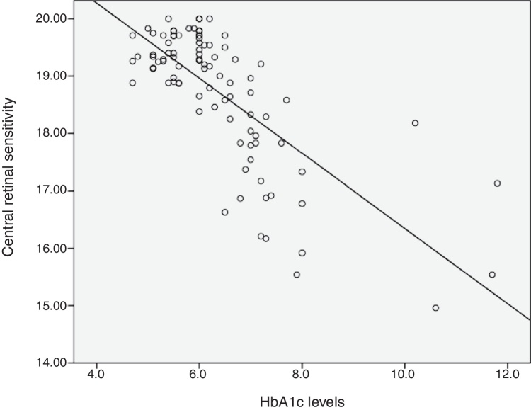
Total mean sensitivity at 10° for group 1 without DR, group 2 with mild NPDR and control group were 18.67±0.83, 17.98±1.42 and 19.45±0.34 (dB), respectively. There was a significant difference in total mean retinal sensitivity at 10° between the 3 groups (F(2,89)=18.14, p=0.001). A simple linear regression was calculated to predict HbA1c based on retinal sensitivity. A significant regression equation was found (F(1,90)=107.61, p=0.0001, with an R of 0.545). The linear regression analysis revealed that there was a 0.64dB decline in mean retinal sensitivity within the central 10° diameter with an increase of 1mmHg of HbA1c.

CONCLUSION

Retinal sensitivity at the central 10° of the macula is affected by changes in HbA1c values.

摘要

目的

本研究旨在使用MP-1微视野计确定糖化血红蛋白(HbA1c)值与中心10°视网膜敏感度之间的关系。

方法

对32名健康受试者(对照组)和60名糖尿病患者进行了一项前瞻性研究。糖尿病患者被分为两组。第1组由30名无糖尿病视网膜病变(DR)的患者组成,第2组有30名轻度非增殖性DR患者。使用MP-1对所有受试者的视网膜中心10°(黄斑)进行全阈值微视野检查,共32个点。使用线性回归分析计算光敏感度与HbA1c值之间的关系。

结果

第1组无DR、第2组轻度NPDR和对照组在10°的总平均敏感度分别为18.67±0.83、17.98±1.42和19.45±0.34(dB)。三组之间在10°的总平均视网膜敏感度上存在显著差异(F(2,89)=18.14,p=0.001)。计算了一个基于视网膜敏感度预测HbA1c的简单线性回归。发现了一个显著的回归方程(F(1,90)=107.61,p=0.0001,R为0.545)。线性回归分析显示,随着HbA1c每增加1mmHg,中心10°直径内的平均视网膜敏感度下降0.64dB。

结论

黄斑中心10°的视网膜敏感度受HbA1c值变化的影响。

https://cdn.ncbi.nlm.nih.gov/pmc/blobs/d309/6612021/5ad329f60c88/gr1.jpg

相似文献

1
HbA1c and retinal sensitivity in diabetics using microperimetry.使用微视野计检测糖尿病患者的糖化血红蛋白(HbA1c)与视网膜敏感度
J Optom. 2019 Jul-Sep;12(3):174-179. doi: 10.1016/j.optom.2018.03.007. Epub 2018 May 26.
2
Structural and Functional Abnormalities in Early-stage Diabetic Retinopathy.早期糖尿病视网膜病变的结构和功能异常。
Curr Eye Res. 2020 Aug;45(8):975-985. doi: 10.1080/02713683.2019.1705983. Epub 2020 Jan 10.
3
Measuring retinal sensitivity with the microperimeter in patients with diabetes.用微视野计测量糖尿病患者的视网膜敏感度。
Retina. 2012 Jul;32(7):1302-9. doi: 10.1097/IAE.0b013e3182365a24.
4
Fundus microperimetry patterns of fixation in type 2 diabetic patients with diffuse macular edema.2型糖尿病合并弥漫性黄斑水肿患者的眼底微视野注视模式
Retina. 2007 Jan;27(1):21-9. doi: 10.1097/01.iae.0000256658.71864.ca.
5
Macular retinal sensitivity using MP-1 in healthy Malaysian subjects of different ages.在不同年龄的健康马来西亚受试者中使用MP-1测量黄斑视网膜敏感度。
J Optom. 2015 Oct-Dec;8(4):266-72. doi: 10.1016/j.optom.2015.04.001. Epub 2015 May 27.
6
Contrast sensitivity is associated with outer-retina thickness in early-stage diabetic retinopathy.对比敏感度与早期糖尿病视网膜病变中外层视网膜厚度有关。
Acta Ophthalmol. 2020 Mar;98(2):e224-e231. doi: 10.1111/aos.14241. Epub 2019 Sep 13.
7
Rod function in diabetic patients without and with early diabetic retinopathy.无早期糖尿病视网膜病变及有早期糖尿病视网膜病变的糖尿病患者的视杆功能。
Eur J Ophthalmol. 2016 Aug 4;26(5):418-24. doi: 10.5301/ejo.5000800. Epub 2016 May 13.
8
[Reduction of retinal light sensitivity in diabetic patients].[糖尿病患者视网膜光敏感度降低]
Klin Monbl Augenheilkd. 2003 Dec;220(12):868-72. doi: 10.1055/s-2003-812556.
9
Comparison of macular retinal sensitivity and its contribution to the foveal sensitivity between diabetic and non-diabetic patients with normal visual acuity.视力正常的糖尿病患者与非糖尿病患者黄斑视网膜敏感性及其对中央凹敏感性的贡献比较。
J Optom. 2019 Jul-Sep;12(3):180-185. doi: 10.1016/j.optom.2018.08.001. Epub 2018 Oct 28.
10
Retinal thickness profile of individuals with diabetes.糖尿病患者的视网膜厚度分布图
Ophthalmic Physiol Opt. 2016 Mar;36(2):158-66. doi: 10.1111/opo.12263. Epub 2015 Dec 21.

引用本文的文献

1
Ocular findings in children, adolescents and young adults with type 1 diabetes mellitus in ghana: a cross-sectional study.加纳1型糖尿病儿童、青少年及青年的眼部检查结果:一项横断面研究
BMC Ophthalmol. 2025 Jul 1;25(1):366. doi: 10.1186/s12886-025-04187-5.
2
Identifying the severity of diabetic retinopathy by visual function measures using both traditional statistical methods and interpretable machine learning: a cross-sectional study.使用传统统计方法和可解释机器学习识别视觉功能测量的糖尿病视网膜病变严重程度:一项横断面研究。
Diabetologia. 2023 Dec;66(12):2250-2260. doi: 10.1007/s00125-023-06005-3. Epub 2023 Sep 19.
3

本文引用的文献

1
Retinal neurodegeneration may precede microvascular changes characteristic of diabetic retinopathy in diabetes mellitus.在糖尿病中,视网膜神经变性可能先于糖尿病视网膜病变的微血管变化出现。
Proc Natl Acad Sci U S A. 2016 May 10;113(19):E2655-64. doi: 10.1073/pnas.1522014113. Epub 2016 Apr 25.
2
Neurodegeneration in Type 2 Diabetes: Evidence From Spectral-Domain Optical Coherence Tomography.2型糖尿病中的神经退行性变:来自频域光学相干断层扫描的证据。
Invest Ophthalmol Vis Sci. 2015 Oct;56(11):6333-8. doi: 10.1167/iovs.15-17334.
3
Macular retinal sensitivity using MP-1 in healthy Malaysian subjects of different ages.
Relationship Between Glycosylated Hemoglobin Levels and Contrast Sensitivity in People with Type 2 Diabetes Mellitus Without Diabetic Retinopathy.
2 型糖尿病患者糖化血红蛋白水平与无糖尿病视网膜病变患者对比敏感度的关系。
Turk J Ophthalmol. 2022 Dec 28;52(6):394-399. doi: 10.4274/tjo.galenos.2022.99602.
4
Microperimetry in hydroxychloroquine macular toxicity.羟氯喹黄斑毒性的微视野检查。
Rom J Ophthalmol. 2021 Jul-Sep;65(3):235-240. doi: 10.22336/rjo.2021.47.
5
Evidence for Structural and Functional Damage of the Inner Retina in Diabetes With No Diabetic Retinopathy.无糖尿病视网膜病变的糖尿病患者视网膜内层结构和功能损伤的证据
Invest Ophthalmol Vis Sci. 2021 Mar 1;62(3):35. doi: 10.1167/iovs.62.3.35.
6
Serum microRNA Levels in Diabetes Mellitus.糖尿病患者的血清微小RNA水平
Diagnostics (Basel). 2021 Feb 11;11(2):284. doi: 10.3390/diagnostics11020284.
7
Comparison of Fundus-Guided Microperimetry and Multifocal Electroretinography for Evaluating Hydroxychloroquine Maculopathy.用于评估羟氯喹黄斑病变的眼底引导微视野检查与多焦视网膜电图的比较
Transl Vis Sci Technol. 2019 Sep 27;8(5):19. doi: 10.1167/tvst.8.5.19. eCollection 2019 Sep.
在不同年龄的健康马来西亚受试者中使用MP-1测量黄斑视网膜敏感度。
J Optom. 2015 Oct-Dec;8(4):266-72. doi: 10.1016/j.optom.2015.04.001. Epub 2015 May 27.
4
Comparison between multifocal electroretinography and microperimetry in age-related macular degeneration.年龄相关性黄斑变性中多焦视网膜电图与微视野检查的比较
Invest Ophthalmol Vis Sci. 2014 Aug 26;55(10):6431-9. doi: 10.1167/iovs.14-14407.
5
Neurodegeneration in the diabetic eye: new insights and therapeutic perspectives.糖尿病眼中的神经退行性变:新的见解和治疗前景。
Trends Endocrinol Metab. 2014 Jan;25(1):23-33. doi: 10.1016/j.tem.2013.09.005. Epub 2013 Nov 1.
6
Fundus-driven perimetry (microperimetry) compared to conventional static automated perimetry: similarities, differences, and clinical applications.眼底驱动的视野检查(微视野检查)与传统的静态自动视野检查相比:相似之处、不同之处和临床应用。
Can J Ophthalmol. 2013 Oct;48(5):358-63. doi: 10.1016/j.jcjo.2013.03.021. Epub 2013 Sep 2.
7
Neurodegeneration in the pathogenesis of diabetic retinopathy: molecular mechanisms and therapeutic implications.糖尿病性视网膜病变发病机制中的神经退行性变:分子机制和治疗意义。
Curr Med Chem. 2013;20(26):3241-50. doi: 10.2174/09298673113209990027.
8
Diabetic retinopathy.糖尿病视网膜病变
N Engl J Med. 2012 Mar 29;366(13):1227-39. doi: 10.1056/NEJMra1005073.
9
Early neurodegeneration in the retina of type 2 diabetic patients.2 型糖尿病患者视网膜的早期神经退行性变。
Invest Ophthalmol Vis Sci. 2012 May 14;53(6):2715-9. doi: 10.1167/iovs.11-8997.
10
Does neuronal damage precede vascular damage in subjects with type 2 diabetes mellitus and having no clinical diabetic retinopathy?2 型糖尿病患者无临床糖尿病视网膜病变时,神经元损伤是否先于血管损伤?
Ophthalmic Res. 2012;47(4):202-7. doi: 10.1159/000333220. Epub 2011 Dec 16.