The First Clinical Medical College, Fujian Medical University, Fuzhou, Fujian, China.
Department of Cardiology, Affiliated Hospital of Putian University, Putian, Fujian, China.
Oxid Med Cell Longev. 2018 Apr 15;2018:1714896. doi: 10.1155/2018/1714896. eCollection 2018.
Upregulation of prolyl isomerase-1 (Pin1) protein expression and activity was associated with the pathogenesis of diabetic vasculopathy through induction of endothelial oxidative stress and inflammation. Moreover, VDR agonist protects against high glucose-induced endothelial apoptosis through the inhibition of oxidative stress. We aimed to explore the effects of the VDR agonist on diabetes-associated endothelial dysfunction and the role of Pin1 in this process.
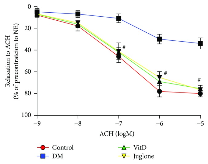
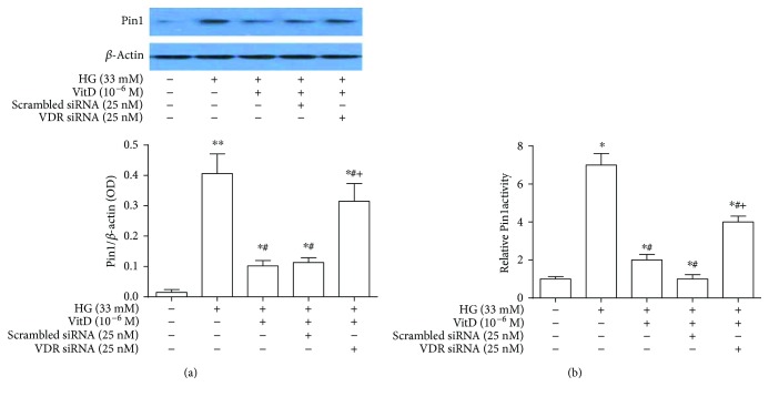
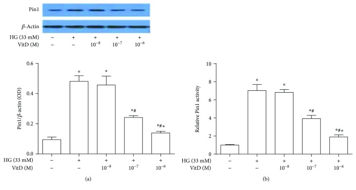
Streptozocin-induced diabetic mice were randomly treated with vehicle, VDR agonist (10 g/kg/d, i.g., twice a week), or Pin1 inhibitor, Juglone (1 mg/kg/d, i.p., every other day), for eight weeks. In parallel, human umbilical vein endothelial cells (HUVECs) exposed to high-glucose condition were treated with 1,25-dihydroxyvitamin D and Juglone or vehicle for 72 hours. Organ chamber experiments were performed to assess endothelium-dependent relaxation to acetylcholine. Circulatory levels of Pin1, SOD, MDA, IL-1, IL-6, and NO in diabetic mice, Pin1 protein expression and activity, subcellular distribution of p66Shc, and NF-B p65 in high glucose-cultured HUVECs were determined.
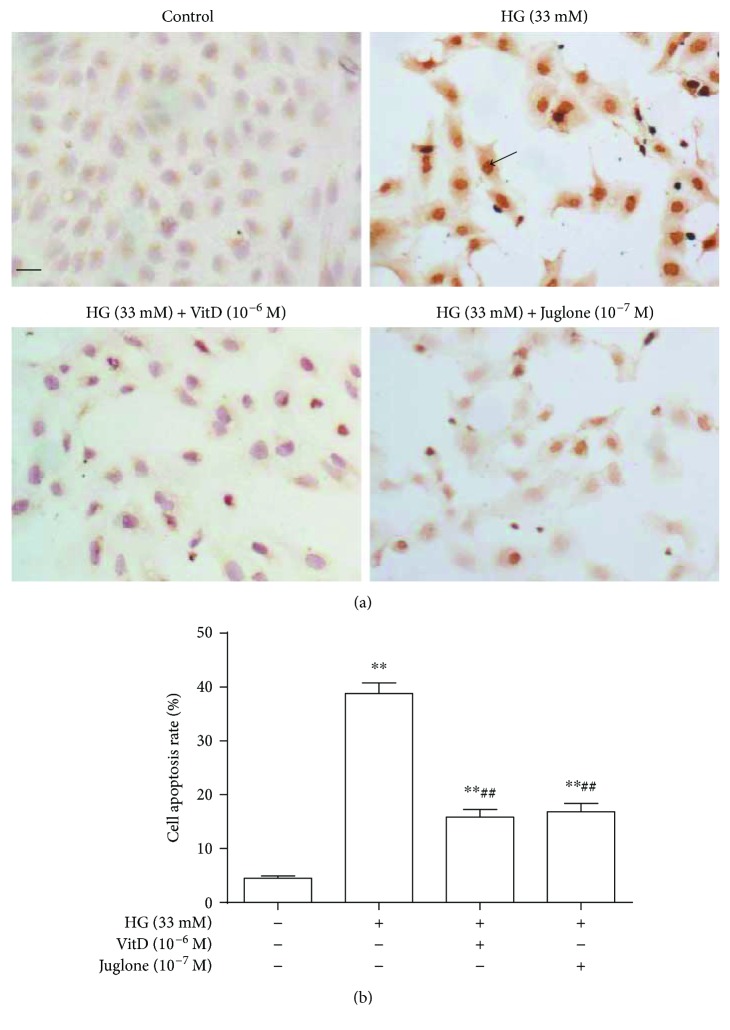
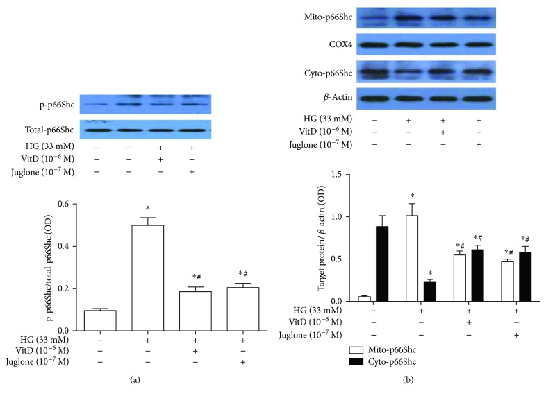
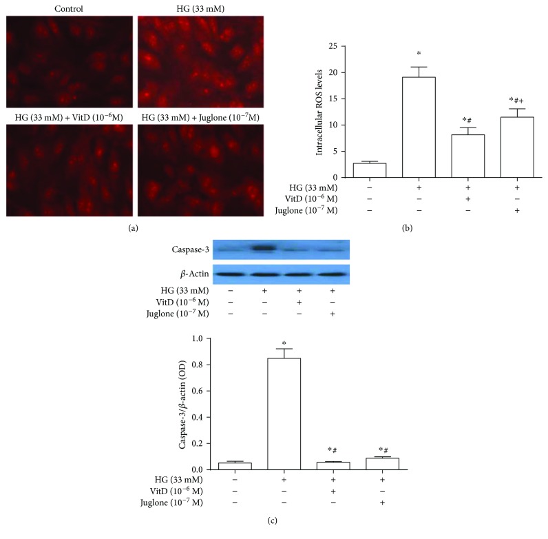
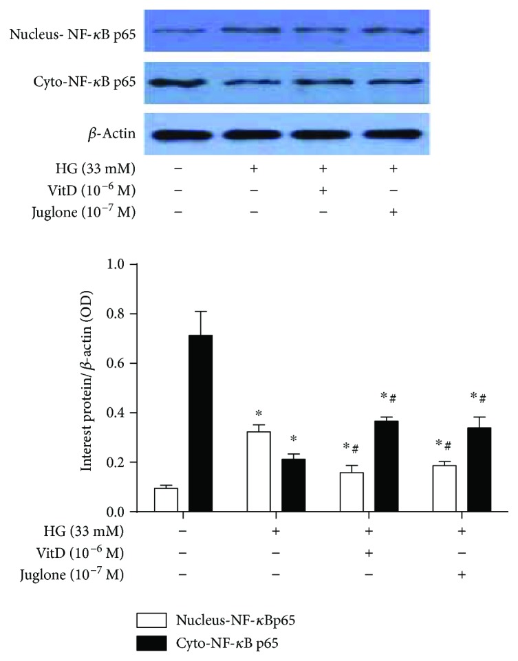
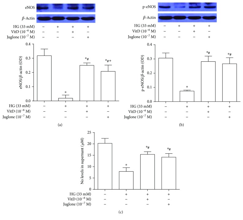
Both VDR agonist and Juglone significantly improved diabetes-associated endothelial dysfunction and reduced high glucose-induced endothelial apoptosis. Mechanistically, the circulatory levels of SOD and NO were increased compared with those of vehicle-treated diabetic mice. Additionally, Pin1 protein expression and activity, p66Shc mitochondrial translocation, and NF-B p65 in high glucose-cultured HUVECs were also inhibited by VDR agonist and Juglone. Knockdown of VDR abolished the inhibitory effects of VDR agonist on high glucose-induced upregulation of Pin1 protein expression and activity.
VDR agonist prevents diabetic endothelial dysfunction through inhibition of Pin1-mediated mitochondrial oxidative stress and inflammation.
脯氨酰基异构酶-1(Pin1)蛋白表达和活性的上调与糖尿病血管病变的发病机制有关,通过诱导内皮氧化应激和炎症。此外,维生素 D 受体激动剂通过抑制氧化应激来保护高葡萄糖诱导的内皮细胞凋亡。我们旨在探讨维生素 D 受体激动剂对糖尿病相关内皮功能障碍的影响,以及 Pin1 在这一过程中的作用。
链脲佐菌素诱导的糖尿病小鼠随机接受载体、维生素 D 受体激动剂(10μg/kg/d,ig,每周两次)或 Pin1 抑制剂 Juglone(1mg/kg/d,ip,每隔一天)治疗 8 周。同时,将人脐静脉内皮细胞(HUVECs)暴露于高葡萄糖环境中,用 1,25-二羟基维生素 D 和 Juglone 或载体处理 72 小时。通过器官室实验评估乙酰胆碱依赖性内皮舒张。测定糖尿病小鼠循环中 Pin1、SOD、MDA、IL-1、IL-6 和 NO 的水平,高葡萄糖培养的 HUVECs 中 Pin1 蛋白表达和活性、p66Shc 的亚细胞分布以及 NF-B p65。
维生素 D 受体激动剂和 Juglone 均显著改善了糖尿病相关的内皮功能障碍,并减少了高葡萄糖诱导的内皮细胞凋亡。机制上,与载体处理的糖尿病小鼠相比,循环中的 SOD 和 NO 水平升高。此外,维生素 D 受体激动剂和 Juglone 还抑制了高葡萄糖培养的 HUVECs 中 Pin1 蛋白表达和活性、p66Shc 的线粒体易位以及 NF-B p65。敲低维生素 D 受体消除了维生素 D 受体激动剂对高葡萄糖诱导的 Pin1 蛋白表达和活性上调的抑制作用。
维生素 D 受体激动剂通过抑制 Pin1 介导的线粒体氧化应激和炎症来预防糖尿病内皮功能障碍。