Department of Otolaryngology-Head and Neck Surgery, Eye, Ear, Nose and Throat Hospital, Fudan University, Shanghai, China.
Key Clinical Disciplines of Otorhinolaryngology of Shanghai, Shanghai, China.
Biomed Res Int. 2018 Apr 17;2018:1230151. doi: 10.1155/2018/1230151. eCollection 2018.
To investigate the potential use of indirect computed tomography lymphography (CT-LG) with dendrimer-entrapped gold nanoparticles (Au DENPs) in the localization and enhanced imaging of cervical sentinel lymph node (SLN) on rabbit model.
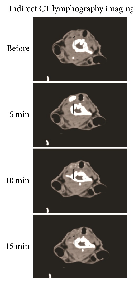
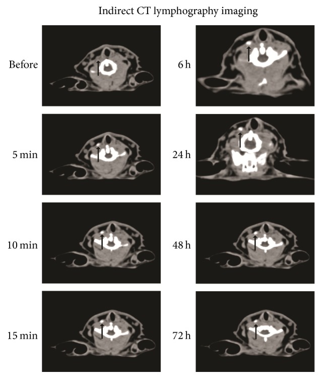
Twelve rabbits were randomly divided into two groups: the positive control group and the experimental group. In the control group, indirect CT-LG was performed with the injection of 0.5 ml activated carbon nanoparticles (ACNP) and Omnipaque mixture suspension in the right tongue submucosa. CT images were acquired before the injection and 1, 5, 10, and 15 min after the injection, respectively. In the experimental group, indirect CT-LG injection with 0.5 ml Au DENPs suspension was performed in the right tongue submucosa. CT images were obtained before the injection and 1, 5, 10, and 15 min and 1, 2, 6, 24, 48, and 72 h after the injection, respectively. Then, SLN identification and enhancement characteristics were evaluated.
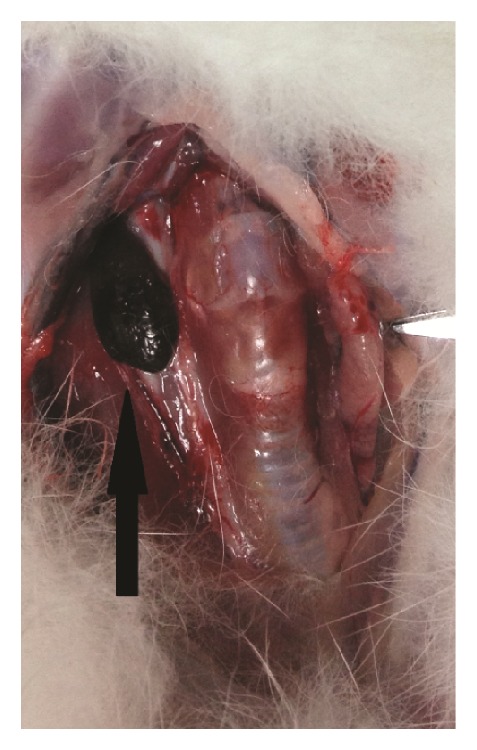
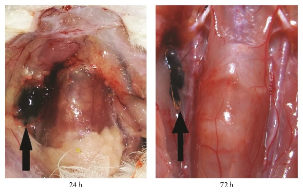
Indirect CT-LG revealed the enhancement of one right deep cervical lymph nodes in all animals, which was SLN. SLN location was marked with black color (ACNP dye) or purple-black color (Au DENPs dye). At each detection time point, the enhanced SLN attenuation values of control rabbits were statistically significantly higher than that of the plain scan, respectively ( < 0.05). Also the values of experimental rabbits were statistically significantly higher than that of the control at the same time point after injection ( < 0.05). The detection rate of SLN was 100%.
Indirect CT-LG with injection of Au DENPs as CT contrast agents can locate the SLN for a long period of time and enrich the SLN black dye. It is helpful for SLNs identification during the operation.
探讨树枝状高分子纳米金包裹物(Au DENPs)间接计算机断层淋巴造影(CT-LG)在兔模型颈哨淋巴结(SLN)定位和增强成像中的潜在应用。
将 12 只兔随机分为两组:阳性对照组和实验组。在对照组中,于右侧舌黏膜下注射 0.5ml 活性炭纳米颗粒(ACNP)和欧乃派克混合悬液,行间接 CT-LG。分别于注射前和注射后 1、5、10 和 15min 采集 CT 图像。实验组于右侧舌黏膜下注射 0.5ml Au DENPs 混悬液,行间接 CT-LG。分别于注射前和注射后 1、5、10、15min 和 1、2、6、24、48 和 72h 采集 CT 图像。评估 SLN 的识别和增强特征。
间接 CT-LG 显示所有动物均有 1 个右侧深部颈淋巴结增强,即 SLN。SLN 的位置用黑色(ACNP 染料)或紫黑色(Au DENPs 染料)标记。在每个检测时间点,对照组兔的增强 SLN 衰减值均明显高于平扫( < 0.05)。且注射后同一时间点实验组兔的增强 SLN 衰减值均明显高于对照组( < 0.05)。SLN 的检出率为 100%。
以 Au DENPs 为 CT 对比剂的间接 CT-LG 可以长时间定位 SLN,并使 SLN 黑染剂富集,有助于手术中识别 SLN。