From the Experimental Atherosclerosis Section, National Heart, Lung, and Blood Institute (X.J., Y.L., J.C., R.F., H.S.K.).
Scanning Probe Microscopy Unit, National Institute of Biomedical Imaging and Bioengineering (E.K.D.).
Arterioscler Thromb Vasc Biol. 2018 Jul;38(7):1504-1518. doi: 10.1161/ATVBAHA.118.311269. Epub 2018 May 31.
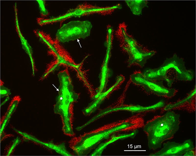
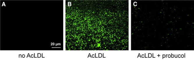
Cells use various mechanisms to maintain cellular cholesterol homeostasis including efflux of cholesterol from the cellular plasma membrane to cholesterol acceptors such as HDLs (high-density lipoproteins). Little is known about the transfer of cholesterol from cells into the extracellular matrix. Using a unique monoclonal antibody that detects ordered cholesterol arrays (ie, cholesterol micro[or nano]-domains), we previously identified that particles containing these cholesterol domains accumulate in the extracellular matrix during cholesterol enrichment of human monocyte-derived macrophages and are found in atherosclerotic lesions. In this study, we further investigate these deposited particles containing cholesterol microdomains and discover their unexpected morphology.
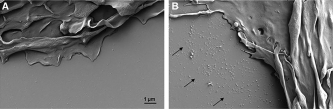
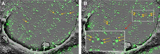
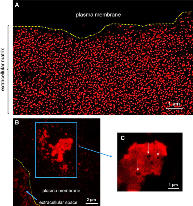
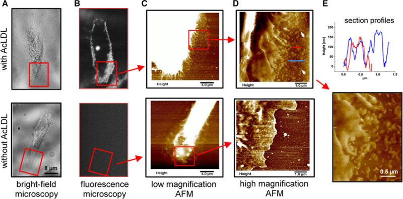
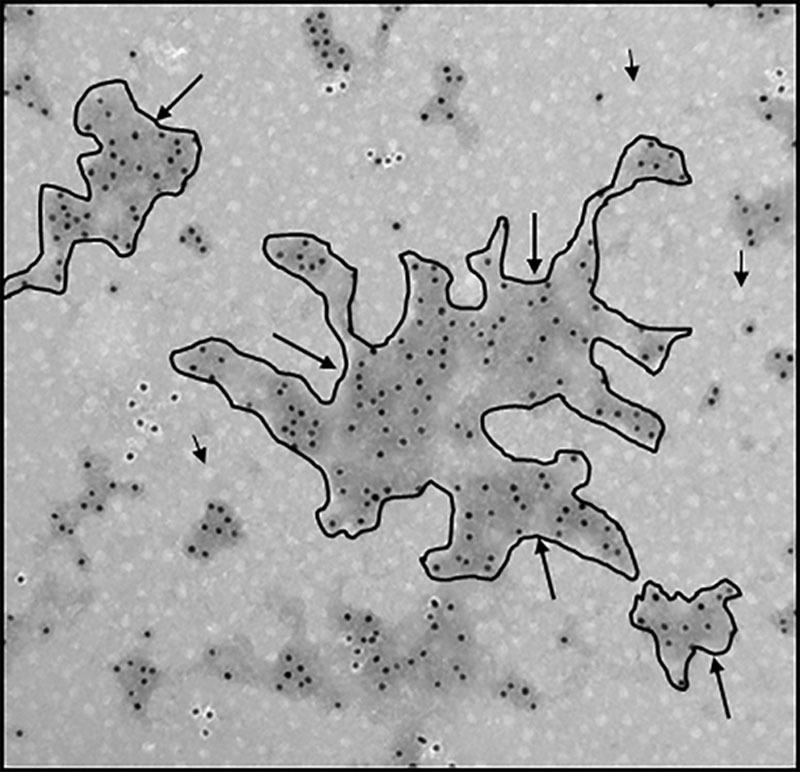
Although appearing spherical at the resolution of the conventional fluorescence microscope, super-resolution immunofluorescence and atomic force microscopy of in situ cholesterol microdomains, and immunoelectron microscopy of isolated cholesterol microdomains revealed that the microdomains are not vesicles or 3-dimensional crystals but rather appear as branching irregularly shaped deposits of varying size. These cholesterol microdomain-containing deposits are shed from the plasma membrane into the extracellular matrix.
To date, research on cellular excretion of excess cholesterol has demonstrated cellular cholesterol efflux in the form of membranous vesicles and discoidal HDL particles released into the fluid-phase medium. Shedding of plasma membrane cholesterol microdomains provides an additional mechanism for cells such as macrophages to maintain plasma membrane cholesterol homeostasis. Furthermore, recognition that macrophages shed cholesterol microdomains into the extracellular matrix is important to our understanding of extracellular buildup of cholesterol in atherosclerosis.
细胞使用各种机制来维持细胞内胆固醇稳态,包括将胆固醇从细胞膜质排出到胆固醇受体,如高密度脂蛋白(HDL)。关于胆固醇从细胞转移到细胞外基质的情况知之甚少。我们使用一种独特的单克隆抗体来检测有序的胆固醇排列(即胆固醇微[或纳米]域),此前发现,在人单核细胞源性巨噬细胞胆固醇富集过程中,含有这些胆固醇域的颗粒会积聚在细胞外基质中,并存在于动脉粥样硬化病变中。在这项研究中,我们进一步研究了这些含有胆固醇微域的沉积颗粒,并发现了它们意想不到的形态。
尽管在常规荧光显微镜的分辨率下呈现球形,但胆固醇微域的超分辨率免疫荧光和原子力显微镜以及分离的胆固醇微域的免疫电子显微镜显示,这些微域不是囊泡或 3 维晶体,而是呈现出分支的不规则形状的沉积物,大小不一。这些含有胆固醇微域的沉积物从质膜脱落到细胞外基质中。
迄今为止,关于细胞过量胆固醇排泄的研究表明,细胞胆固醇以膜性小泡和释放到液相介质中的碟形 HDL 颗粒的形式流出。质膜胆固醇微域的脱落为巨噬细胞等细胞提供了一种维持质膜胆固醇稳态的额外机制。此外,认识到巨噬细胞将胆固醇微域脱落到细胞外基质中,对于我们理解动脉粥样硬化中胆固醇在细胞外的积累非常重要。