• 文献检索
  • 文档翻译
  • 深度研究
  • 学术资讯
  • Suppr Zotero 插件Zotero 插件
  • 邀请有礼
  • 套餐&价格
  • 历史记录
应用&插件
Suppr Zotero 插件Zotero 插件浏览器插件Mac 客户端Windows 客户端微信小程序
定价
高级版会员购买积分包购买API积分包
服务
文献检索文档翻译深度研究API 文档MCP 服务
关于我们
关于 Suppr公司介绍联系我们用户协议隐私条款
关注我们

Suppr 超能文献

核心技术专利:CN118964589B侵权必究
粤ICP备2023148730 号-1Suppr @ 2026

文献检索

告别复杂PubMed语法,用中文像聊天一样搜索,搜遍4000万医学文献。AI智能推荐,让科研检索更轻松。

立即免费搜索

文件翻译

保留排版,准确专业,支持PDF/Word/PPT等文件格式,支持 12+语言互译。

免费翻译文档

深度研究

AI帮你快速写综述,25分钟生成高质量综述,智能提取关键信息,辅助科研写作。

立即免费体验

颅颈区解剖:影像学表现、陷阱及模仿疾病:图文综述

Craniocervical Dissections: Radiologic Findings, Pitfalls, Mimicking Diseases: A Pictorial Review.

作者信息

Mehdi Elnur, Aralasmak Ayse, Toprak Huseyin, Yıldız Seyma, Kurtcan Serpil, Kolukisa Mehmet, Asıl Talip, Alkan Alpay

机构信息

Bezmialem Vakif University, Department of Radiology, Istanbul, Turkey.

Bezmialem Vakif University, Department of Neurology, Istanbul, Turkey.

出版信息

Curr Med Imaging Rev. 2018 Apr;14(2):207-222. doi: 10.2174/1573405613666170403102235.

DOI:10.2174/1573405613666170403102235
PMID:29853818
原文链接:https://pmc.ncbi.nlm.nih.gov/articles/PMC5902863/
Abstract

BACKGROUND

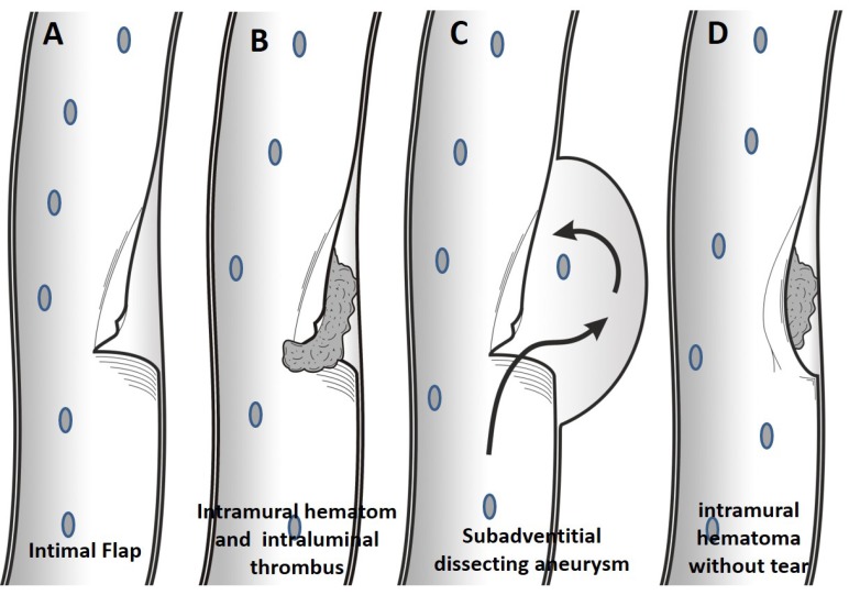
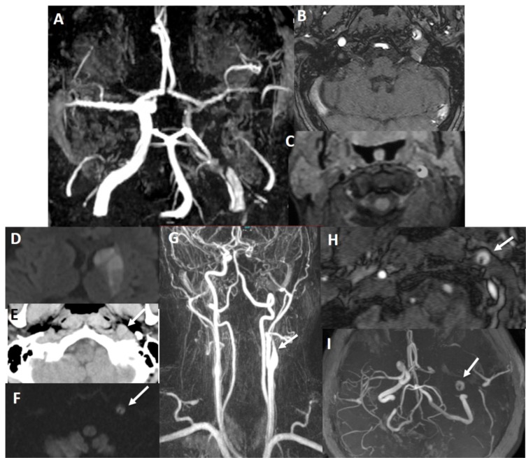
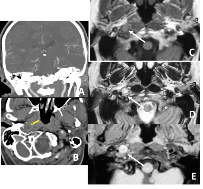
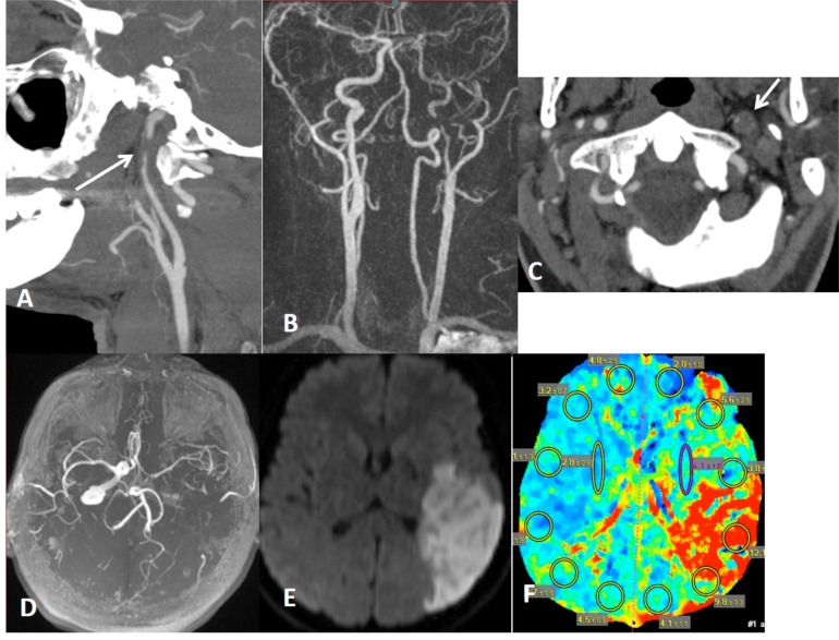
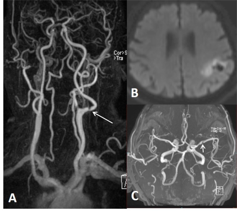
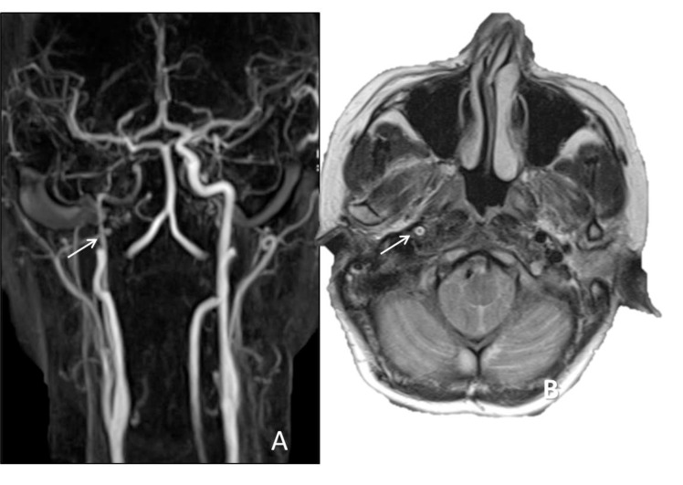
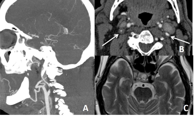
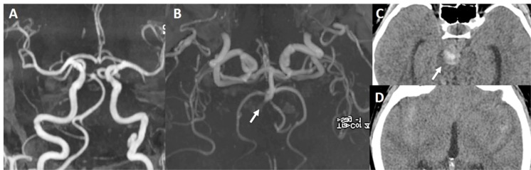
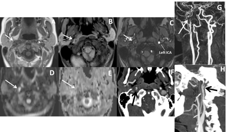
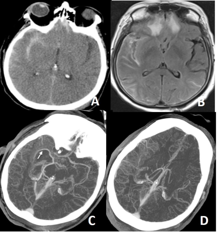
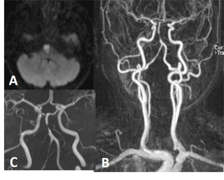
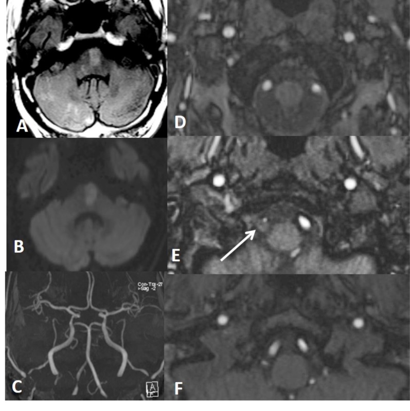
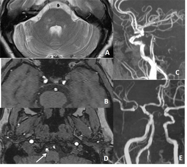
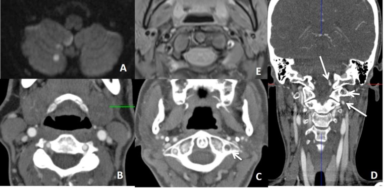
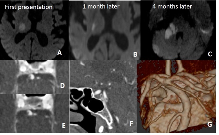
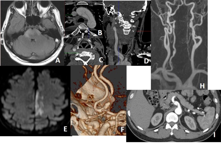
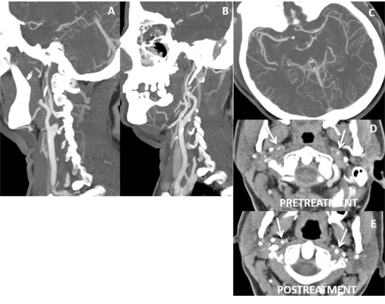
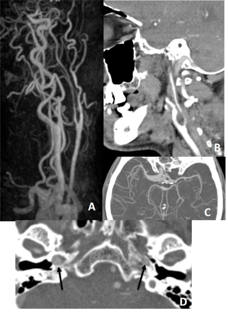
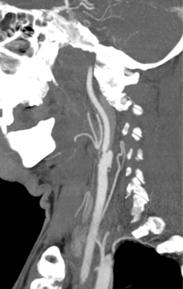
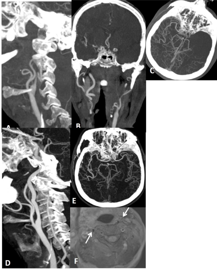
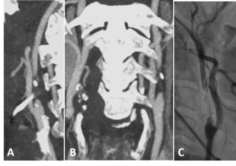
Craniocervical Dissections (CCD) are a crucial emergency state causing 20% of strokes in patients under the age of 45. Although DSA (digital substraction angiography) is regarded as the gold standard, noninvasive methods of CT, CTA and MRI, MRA are widely used for diagnosis.

AIM

Our aim is to illustrate noninvasive imaging findings in CCD.

CONCLUSION

Emphasizing on diagnostic pitfalls, limitations and mimicking diseases.

摘要

背景

颅颈夹层分离(CCD)是一种关键的紧急状态,在45岁以下患者中导致20%的中风。尽管数字减影血管造影(DSA)被视为金标准,但CT、CTA以及MRI、MRA等非侵入性方法也广泛用于诊断。

目的

我们的目的是阐述CCD的非侵入性影像学表现。

结论

强调诊断陷阱、局限性及相似疾病。

https://cdn.ncbi.nlm.nih.gov/pmc/blobs/1e87/5902863/024fb5124961/CMIR-14-207_F22.jpg
https://cdn.ncbi.nlm.nih.gov/pmc/blobs/1e87/5902863/37b6c3ff2bc4/CMIR-14-207_F1.jpg
https://cdn.ncbi.nlm.nih.gov/pmc/blobs/1e87/5902863/9b87f5e82d30/CMIR-14-207_F2.jpg
https://cdn.ncbi.nlm.nih.gov/pmc/blobs/1e87/5902863/95ef9c5a6879/CMIR-14-207_F3.jpg
https://cdn.ncbi.nlm.nih.gov/pmc/blobs/1e87/5902863/32a63e7cef2a/CMIR-14-207_F4.jpg
https://cdn.ncbi.nlm.nih.gov/pmc/blobs/1e87/5902863/74038f97c5ad/CMIR-14-207_F5.jpg
https://cdn.ncbi.nlm.nih.gov/pmc/blobs/1e87/5902863/0b0d0bbd2ac2/CMIR-14-207_F6.jpg
https://cdn.ncbi.nlm.nih.gov/pmc/blobs/1e87/5902863/17928176a030/CMIR-14-207_F7.jpg
https://cdn.ncbi.nlm.nih.gov/pmc/blobs/1e87/5902863/e398e4ae1e38/CMIR-14-207_F8.jpg
https://cdn.ncbi.nlm.nih.gov/pmc/blobs/1e87/5902863/f4405a3d56d9/CMIR-14-207_F9.jpg
https://cdn.ncbi.nlm.nih.gov/pmc/blobs/1e87/5902863/682a3d424ab2/CMIR-14-207_F10.jpg
https://cdn.ncbi.nlm.nih.gov/pmc/blobs/1e87/5902863/ab7daf4ef531/CMIR-14-207_F11.jpg
https://cdn.ncbi.nlm.nih.gov/pmc/blobs/1e87/5902863/a15d6cf5c5e1/CMIR-14-207_F12.jpg
https://cdn.ncbi.nlm.nih.gov/pmc/blobs/1e87/5902863/18d09a0d65cd/CMIR-14-207_F13.jpg
https://cdn.ncbi.nlm.nih.gov/pmc/blobs/1e87/5902863/add2e4b5e8f3/CMIR-14-207_F14.jpg
https://cdn.ncbi.nlm.nih.gov/pmc/blobs/1e87/5902863/9ac35f433436/CMIR-14-207_F15.jpg
https://cdn.ncbi.nlm.nih.gov/pmc/blobs/1e87/5902863/06ac79cdaab2/CMIR-14-207_F16.jpg
https://cdn.ncbi.nlm.nih.gov/pmc/blobs/1e87/5902863/67ec7eb72637/CMIR-14-207_F17.jpg
https://cdn.ncbi.nlm.nih.gov/pmc/blobs/1e87/5902863/b43ddd3c13d6/CMIR-14-207_F18.jpg
https://cdn.ncbi.nlm.nih.gov/pmc/blobs/1e87/5902863/5bdfa59efc58/CMIR-14-207_F19.jpg
https://cdn.ncbi.nlm.nih.gov/pmc/blobs/1e87/5902863/0c07002f389e/CMIR-14-207_F20.jpg
https://cdn.ncbi.nlm.nih.gov/pmc/blobs/1e87/5902863/535909ade7f4/CMIR-14-207_F21.jpg
https://cdn.ncbi.nlm.nih.gov/pmc/blobs/1e87/5902863/024fb5124961/CMIR-14-207_F22.jpg

相似文献

1
Craniocervical Dissections: Radiologic Findings, Pitfalls, Mimicking Diseases: A Pictorial Review.颅颈区解剖:影像学表现、陷阱及模仿疾病:图文综述
Curr Med Imaging Rev. 2018 Apr;14(2):207-222. doi: 10.2174/1573405613666170403102235.
2
Comparison of multidetector CT angiography and MR imaging of cervical artery dissection.多层螺旋CT血管造影与磁共振成像对颈动脉夹层的比较。
AJNR Am J Neuroradiol. 2008 Oct;29(9):1753-60. doi: 10.3174/ajnr.A1189. Epub 2008 Jul 17.
3
Combination of noninvasive neurovascular imaging modalities in stroke patients: patterns of use and impact on need for digital subtraction angiography.联合应用无创性神经血管影像学检查手段评估脑卒中患者:使用模式及其对数字减影血管造影需求的影响。
J Stroke Cerebrovasc Dis. 2013 Oct;22(7):e53-8. doi: 10.1016/j.jstrokecerebrovasdis.2012.03.020. Epub 2012 May 11.
4
Craniocervical arterial dissection in children: diagnosis and treatment.儿童颅颈动脉夹层:诊断与治疗。
Curr Treat Options Neurol. 2011 Dec;13(6):636-48. doi: 10.1007/s11940-011-0149-2.
5
CT versus MR Techniques in the Detection of Cervical Artery Dissection.CT与MR技术在检测颈内动脉夹层中的应用比较
J Neuroimaging. 2017 Nov;27(6):607-612. doi: 10.1111/jon.12451. Epub 2017 Jun 2.
6
Diagnostic pitfalls in postinterventional intraarterial magnetic resonance angiography after recanalization of femoropopliteal arterial occlusions.股腘动脉闭塞再通术后介入性动脉内磁共振血管造影的诊断陷阱
Acta Radiol. 2008 Dec;49(10):1129-36. doi: 10.1080/02841850802508942.
7
Surveillance imaging after intracranial stent implantation: non-invasive imaging compared with digital subtraction angiography.颅内支架植入术后的监测成像:非侵入性成像与数字减影血管造影比较。
J Neurointerv Surg. 2013 Jul;5(4):361-5. doi: 10.1136/neurintsurg-2012-010341. Epub 2012 May 28.
8
High-resolution 3D volumetric contrast-enhanced MR angiography with a blood pool agent (ferumoxytol) for diagnostic evaluation of pediatric brain arteriovenous malformations.使用血池造影剂(铁羧麦芽糖)的高分辨率3D容积对比增强磁共振血管造影术用于小儿脑动静脉畸形的诊断评估。
J Neurosurg Pediatr. 2018 Sep;22(3):251-260. doi: 10.3171/2018.3.PEDS17723. Epub 2018 Jun 8.
9
Imaging of Carotid Dissection.颈动脉夹层的影像学表现。
Curr Pain Headache Rep. 2019 Jan 19;23(1):2. doi: 10.1007/s11916-019-0741-9.
10
Imaging modalities for renal artery stenosis in suspected renovascular hypertension: prospective intraindividual comparison of color Doppler US, CT angiography, GD-enhanced MR angiography, and digital substraction angiography.疑似肾血管性高血压患者肾动脉狭窄的成像方式:彩色多普勒超声、CT血管造影、钆增强磁共振血管造影和数字减影血管造影的前瞻性个体内比较
Ren Fail. 2007;29(3):295-302. doi: 10.1080/08860220601166305.

引用本文的文献

1
Neuroimaging for neurovascular complications of traumatic brain injury.创伤性脑损伤神经血管并发症的神经影像学检查
Crit Care. 2025 Aug 18;29(1):362. doi: 10.1186/s13054-025-05586-2.
2
Outer lumen arterial imaging with CTA: A potential tool to diagnose vertebral artery dissection.CTA 进行的外腔动脉成像:诊断椎动脉夹层的潜在工具。
Radiol Case Rep. 2025 Apr 19;20(7):3376-3379. doi: 10.1016/j.radcr.2025.03.041. eCollection 2025 Jul.
3
Predicting vessel recanalization in extracranial internal carotid artery dissection: a nomogram based on ultrasonography and clinical features.

本文引用的文献

1
In Vivo Validation of Simultaneous Non-Contrast Angiography and intraPlaque Hemorrhage (SNAP) Magnetic Resonance Angiography: An Intracranial Artery Study.同时进行非对比血管造影和斑块内出血(SNAP)磁共振血管造影的体内验证:一项颅内动脉研究。
PLoS One. 2016 Feb 10;11(2):e0149130. doi: 10.1371/journal.pone.0149130. eCollection 2016.
2
Clinical Characteristics of Intracranial Reversed Vertebral Artery Flow Evaluated by Transcranial Color Flow Imaging.经颅彩色血流成像评估颅内椎动脉血流逆转的临床特征
J Stroke Cerebrovasc Dis. 2015 Aug;24(8):1775-80. doi: 10.1016/j.jstrokecerebrovasdis.2015.03.044. Epub 2015 Jun 3.
3
预测颅外颈内动脉夹层的血管再通:基于超声检查和临床特征的列线图
Front Neurol. 2025 Apr 7;16:1498182. doi: 10.3389/fneur.2025.1498182. eCollection 2025.
4
Isolated Hypoglossal Nerve Palsy in the Setting of Concurrent Vertebral Artery Dissection and Internal Carotid Artery Dissection Plus Pseudoaneurysm: Case Report and Literature Review.椎动脉夹层和颈内动脉夹层伴假性动脉瘤并发孤立性舌下神经麻痹:病例报告及文献综述
Brain Sci. 2025 Feb 21;15(3):225. doi: 10.3390/brainsci15030225.
5
A Visualized Nomogram to Predict the Risk of Acute Ischemic Stroke Among Patients With Cervical Artery Dissection.一种用于预测颈动脉夹层患者急性缺血性卒中风险的可视化列线图。
Int J Gen Med. 2025 Mar 19;18:1569-1580. doi: 10.2147/IJGM.S507043. eCollection 2025.
6
Spontaneous bilateral vertebral artery dissection complicated by hemodynamic posterior circulation stroke: A case report.自发性双侧椎动脉夹层伴血流动力学性后循环卒中:一例报告
Radiol Case Rep. 2024 Dec 20;20(3):1447-1451. doi: 10.1016/j.radcr.2024.11.095. eCollection 2025 Mar.
7
Predicting outcomes of unruptured intracranial artery dissection with clear symptoms onset using clinical and radiological features.利用临床和影像学特征预测症状明确的未破裂颅内动脉夹层的转归。
Sci Rep. 2024 Oct 1;14(1):22777. doi: 10.1038/s41598-024-73418-4.
8
Association of diabetes and white blood cell count with stroke in patients with carotid artery dissection.糖尿病和白细胞计数与颈动脉夹层患者中风的关系。
BMC Neurol. 2024 Sep 17;24(1):350. doi: 10.1186/s12883-024-03856-0.
9
Dissecting and fusiform aneurysms of the superior cerebellar artery: anatomy, clinical presentation, and treatment outcomes.小脑上动脉夹层及梭形动脉瘤:解剖、临床表现及治疗结果。
Neurosurg Rev. 2024 Aug 31;47(1):516. doi: 10.1007/s10143-024-02734-0.
10
Comparative meta-analysis of endovascular strategies for intracranial dissecting aneurysms: Flow diverters versus stents with or without coiling.颅内夹层动脉瘤血管内治疗策略的比较荟萃分析:血流导向装置与带或不带弹簧圈栓塞的支架对比
Interv Neuroradiol. 2024 Jun 14:15910199241262070. doi: 10.1177/15910199241262070.
Characterization of Craniocervical Artery Dissection by Simultaneous MR Noncontrast Angiography and Intraplaque Hemorrhage Imaging at 3T.
3T磁共振非对比血管造影和斑块内出血成像同步用于颅颈动脉夹层的特征分析
AJNR Am J Neuroradiol. 2015 Sep;36(9):1769-75. doi: 10.3174/ajnr.A4348. Epub 2015 Jun 4.
4
Pathophysiology and risk factors of cervical artery dissection: what have we learnt from large hospital-based cohorts?颈内动脉夹层的病理生理学和危险因素:我们从大型基于医院的队列研究中学到了什么?
Curr Opin Neurol. 2014 Feb;27(1):20-8. doi: 10.1097/WCO.0000000000000056.
5
Clinical characteristics of symptomatic vertebral artery dissection: a systematic review.症状性椎动脉夹层的临床特征:一项系统评价
Neurologist. 2012 Sep;18(5):245-54. doi: 10.1097/NRL.0b013e31826754e1.
6
Cervical carotid artery dissection is associated with styloid process length.颈总动脉夹层与茎突长度有关。
Neurology. 2011 Dec 6;77(23):2061-6. doi: 10.1212/WNL.0b013e31823b4729. Epub 2011 Nov 23.
7
Craniocervical arterial dissection in children: diagnosis and treatment.儿童颅颈动脉夹层:诊断与治疗。
Curr Treat Options Neurol. 2011 Dec;13(6):636-48. doi: 10.1007/s11940-011-0149-2.
8
2011 ASA/ACCF/AHA/AANN/AANS/ACR/ASNR/CNS/SAIP/SCAI/SIR/SNIS/SVM/SVS guideline on the management of patients with extracranial carotid and vertebral artery disease: executive summary.2011年美国麻醉医师协会/美国心脏病学会基金会/美国心脏协会/美国神经病学护士协会/美国神经外科医师协会/美国放射学会/美国神经放射学会/美国神经外科医师协会/介入放射学会/心血管造影和介入学会/放射学会/神经介入学会/血管医学学会/血管外科学会关于颅外颈动脉和椎动脉疾病患者管理的指南:执行摘要
Stroke. 2011 Aug;42(8):e420-63. doi: 10.1161/STR.0b013e3182112d08. Epub 2011 Jan 31.
9
Causes of misinterpretation of cross-sectional imaging studies for dissection of the craniocervical arteries.颅颈动脉夹层的横断面影像学研究解读错误的原因。
AJR Am J Roentgenol. 2011 Jan;196(1):45-52. doi: 10.2214/AJR.10.5384.
10
Cerebrovascular dissections--a review part I: Spontaneous dissections.脑血管夹层——综述部分 I:自发性夹层。
Neurosurgery. 2011 Jan;68(1):242-57; discussion 257. doi: 10.1227/NEU.0b013e3182012323.