Biomedical Electronics Robotics & Devices (BERD) Group, Lab of Medical Physics, School of Medicine, Faculty of Health Sciences, Aristotle University of Thessaloniki (AUTH), 54124 Thessaloniki, Greece.
1st Department of Neurosurgery, "AHEPA" University General Hospital, Aristotle University of Thessaloniki (AUTH), 54636 Thessaloniki, Greece.
Neural Plast. 2018 May 2;2018:9354207. doi: 10.1155/2018/9354207. eCollection 2018.
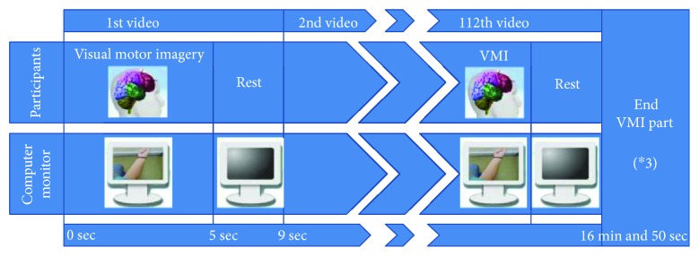
Reciprocal communication of the central and peripheral nervous systems is compromised during spinal cord injury due to neurotrauma of ascending and descending pathways. Changes in brain organization after spinal cord injury have been associated with differences in prognosis. Changes in functional connectivity may also serve as injury biomarkers. Most studies on functional connectivity have focused on chronic complete injury or resting-state condition. In our study, ten right-handed patients with incomplete spinal cord injury and ten age- and gender-matched healthy controls performed multiple visual motor imagery tasks of upper extremities and walking under high-resolution electroencephalography recording. Directed transfer function was used to study connectivity at the cortical source space between sensorimotor nodes. Chronic disruption of reciprocal communication in incomplete injury could result in permanent significant decrease of connectivity in a subset of the sensorimotor network, regardless of positive or negative neurological outcome. Cingulate motor areas consistently contributed the larger outflow (right) and received the higher inflow (left) among all nodes, across all motor imagery categories, in both groups. Injured subjects had higher outflow from left cingulate than healthy subjects and higher inflow in right cingulate than healthy subjects. Alpha networks were less dense, showing less integration and more segregation than beta networks. Spinal cord injury patients showed signs of increased local processing as adaptive mechanism. This trial is registered with NCT02443558.
中枢神经系统和周围神经系统之间的相互交流在脊髓损伤时由于上升和下降通路的神经损伤而受到损害。脊髓损伤后的大脑组织变化与预后的差异有关。功能连接的变化也可以作为损伤的生物标志物。大多数关于功能连接的研究都集中在慢性完全性损伤或静息状态。在我们的研究中,10 名右侧上肢不完全性脊髓损伤患者和 10 名年龄和性别匹配的健康对照者在高分辨率脑电图记录下进行了多次上肢和行走的视觉运动想象任务。定向传递函数用于研究感觉运动节点之间皮质源空间的连通性。不完全损伤中相互交流的慢性中断可能导致感觉运动网络的一部分连接永久性显著下降,无论神经预后是阳性还是阴性。在所有运动想象类别中,两组的所有节点中,扣带回运动区始终贡献较大的输出(右侧),接收较高的输入(左侧)。受伤者的左扣带回比健康者的输出更高,右扣带回比健康者的输入更高。与β网络相比,α网络密度较低,整合度较低,分离度较高。脊髓损伤患者表现出增加局部处理的迹象,作为一种适应机制。本试验在 NCT02443558 注册。