Best Emanuelle J, O'Brien Cecelia M, Carseldine Wendy, Deshpande Aniruddh, Glover Rebecca, Park Felicity
Maternity and Gynaecology, John Hunter Hospital, New Lambton Heights, NSW, Australia.
Maternal Fetal Medicine Unit, John Hunter Hospital, New Lambton Heights, NSW, Australia.
Case Rep Obstet Gynecol. 2018 May 3;2018:5312179. doi: 10.1155/2018/5312179. eCollection 2018.
Fetal volvulus is a rare, yet life-threatening condition that requires skilful diagnosis and management. Volvulus occurs when bowel loops become twisted and the twisting of the mesenteric artery leads to congestion, impaired venous return, and bowel necrosis.
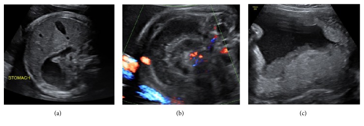
We present a case of fetal ileal volvulus suspected on third trimester ultrasound, complicated by premature labour, small bowel necrosis, and meconium peritonitis. Progressive dilatation and decreased peristalsis of echogenic bowel were noted in the early part of the third trimester. Daily surveillance ultrasound was performed and spontaneous labour occurred at 32 weeks' gestation. A proactive postnatal approach guided by prenatal sonographic findings allowed prompt treatment and an urgent laparotomy was performed for an ileal volvulus with necrosis and meconium peritonitis. A segment of small bowel volvulus was resected and an end-to-end anastomosis was performed with uneventful recovery.
Clinically signs of fetal midgut volvulus are not pathognomonic, such as intestinal dilatation, abdominal mass, ascites, peritoneal calcifications, or polyhydramnios; thus, the diagnosis is often challenging. Complications reported in the literature include perforation and haemorrhagic ascites, which may lead to anaemia, hypovolemia, heart failure, and fetal demise.
This case highlights the importance of assessing the fetal bowel as a part of routine third trimester ultrasound. The case describes the complexity of diagnosis in the fetus, important considerations along with multidisciplinary team approach to management.
胎儿肠扭转是一种罕见但危及生命的疾病,需要熟练的诊断和处理。当肠袢发生扭转且肠系膜动脉扭转导致充血、静脉回流受损和肠坏死时,就会发生肠扭转。
我们报告一例在孕晚期超声检查时怀疑为胎儿回肠扭转的病例,该病例并发早产、小肠坏死和胎粪性腹膜炎。在孕晚期早期,观察到强回声肠管逐渐扩张且蠕动减弱。每日进行超声监测,孕32周时出现自然分娩。根据产前超声检查结果采取的积极产后处理方法使得能够及时进行治疗,针对伴有坏死和胎粪性腹膜炎的回肠扭转紧急实施了剖腹手术。切除了一段小肠扭转肠段并进行了端端吻合,术后恢复顺利。
胎儿中肠扭转的临床体征并无特异性,如肠管扩张、腹部肿块、腹水、腹膜钙化或羊水过多等;因此,诊断往往具有挑战性。文献报道的并发症包括穿孔和出血性腹水,这可能导致贫血、血容量不足、心力衰竭和胎儿死亡。
本病例强调了在孕晚期常规超声检查中评估胎儿肠道的重要性。该病例描述了胎儿诊断的复杂性、重要的注意事项以及多学科团队的处理方法。