Departamento de Fisiología, Biofísica y Neurociencias, Av. Instituto Politécnico Nacional No. 2508, San Pedro Zacatenco, Centro de Investigación y de Estudios Avanzados, 07360 Ciudad de México, Mexico.
Facultad de Ciencias Químicas, 14 Sur y Avenida San Claudio, Benemérita Universidad Autónoma de Puebla, 72570 Puebla, PUE, Mexico.
J Immunol Res. 2018 May 8;2018:1838921. doi: 10.1155/2018/1838921. eCollection 2018.
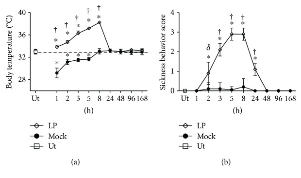
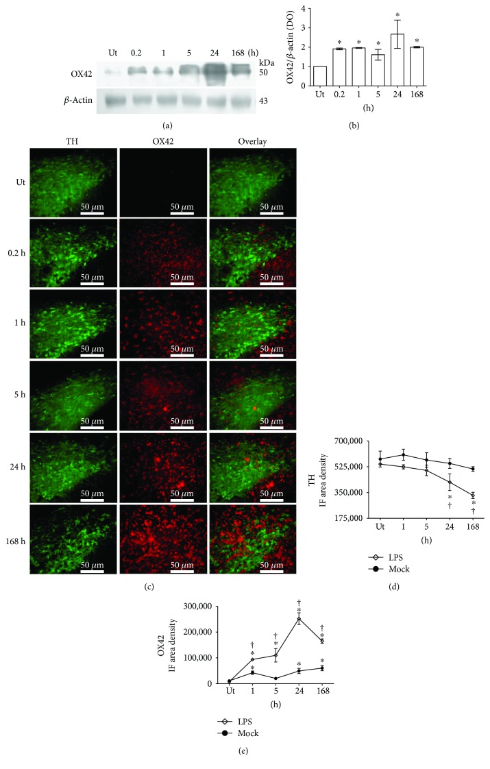
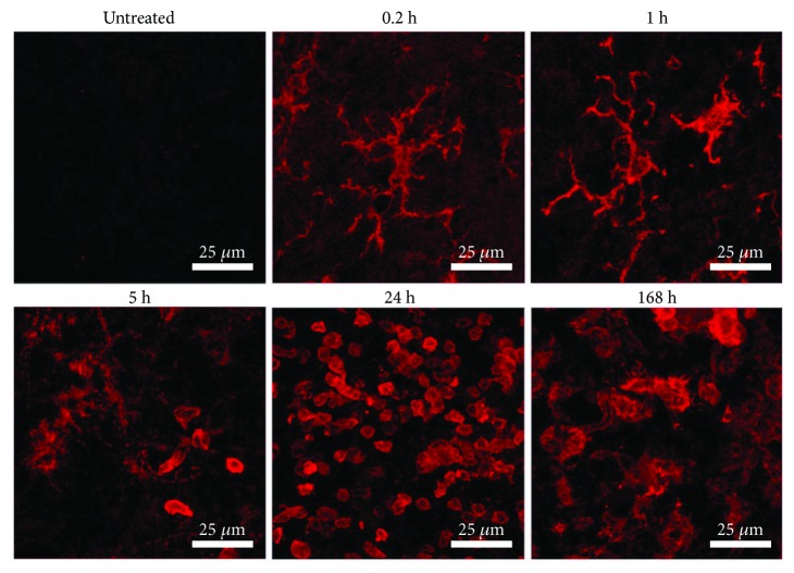
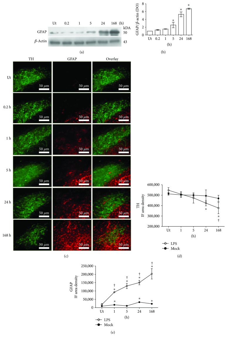
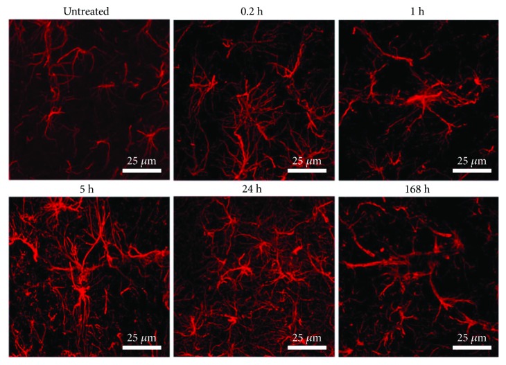
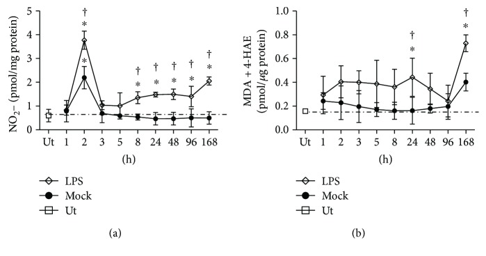
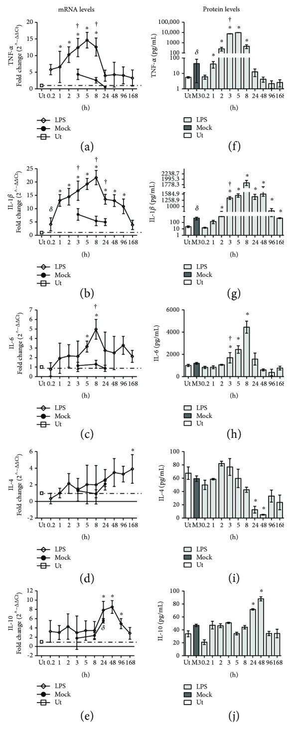
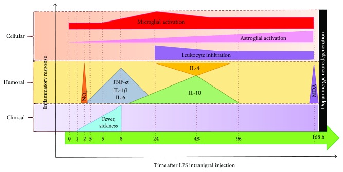
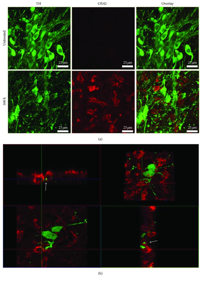
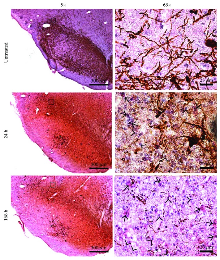
Models of Parkinson's disease with neurotoxins have shown that microglial activation does not evoke a typical inflammatory response in the substantia nigra, questioning whether neuroinflammation leads to neurodegeneration. To address this issue, the archetypal inflammatory stimulus, lipopolysaccharide (LPS), was injected into the rat substantia nigra. LPS induced fever, sickness behavior, and microglial activation (OX42 immunoreactivity), followed by astrocyte activation and leukocyte infiltration (GFAP and CD45 immunoreactivities). During the acute phase of neuroinflammation, pro- and anti-inflammatory cytokines (TNF-, IL-1, IL-6, IL-4, and IL-10) responded differentially at mRNA and protein level. Increased NO production and lipid peroxidation occurred at 168 h after LPS injection. At this time, evidence of neurodegeneration could be seen, entailing decreased tyrosine hydroxylase (TH) immunoreactivity, irregular body contour, and prolongation discontinuity of TH cells, as well as apparent phagocytosis of TH cells by OX42 cells. Altogether, these results show that LPS evokes a typical inflammatory response in the substantia nigra that is followed by dopaminergic neurodegeneration.
帕金森病模型用神经毒素表明,小胶质细胞的激活不会引起黑质中典型的炎症反应,这质疑了神经炎症是否会导致神经退行性变。为了解决这个问题,将典型的炎症刺激物脂多糖 (LPS) 注射到大鼠黑质中。LPS 诱导发热、疾病行为和小胶质细胞激活(OX42 免疫反应),随后是星形胶质细胞激活和白细胞浸润(GFAP 和 CD45 免疫反应)。在神经炎症的急性期,促炎和抗炎细胞因子(TNF-α、IL-1、IL-6、IL-4 和 IL-10)在 mRNA 和蛋白质水平上表现出不同的反应。在 LPS 注射后 168 小时,NO 产生和脂质过氧化增加。此时,可以看到神经退行性变的证据,包括酪氨酸羟化酶 (TH) 免疫反应性降低、不规则的身体轮廓和 TH 细胞的连续性中断,以及 TH 细胞被 OX42 细胞明显吞噬。总的来说,这些结果表明 LPS 会在黑质中引发典型的炎症反应,随后是多巴胺能神经退行性变。