Liu Meilin, Chen Xiaolong, Liu Henan, Di Yu
Department of Ophthalmology, Shengjing Affiliated Hospital, China Medical University, Shenyang, Liaoning, People's Republic of China.
Drug Des Devel Ther. 2018 May 21;12:1337-1346. doi: 10.2147/DDDT.S149594. eCollection 2018.
The aim of the study was to investigate the signal transduction mechanism of Hedgehog-vascular endothelial growth factor in oxygen-induced retinopathy (OIR) and the effects of cyclopamine on OIR.
An OIR model was established in C57BL/6J mice exposed to hyperoxia. Two hundred mice were randomly divided into a control group, an OIR group, an OIR-control group (treated with isometric phosphate-buffered saline by intravitreal injection), and a cyclopamine group (treated with cyclopamine by intravitreal injection), with 50 mice in each group. The retinal vascular morphology was observed using adenosine diphosphatase and number counting using hematoxylin and eosin-stained image. Quantitative real-time quantitative polymerase chain reaction was used to detect mRNA expression. Protein location and expression were evaluated using immunohistochemistry and Western blot.
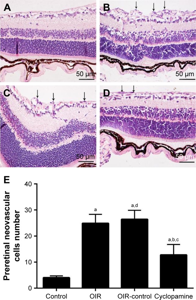
The OIR group and OIR-control group demonstrated large-area pathological neovascularization and nonperfused area when compared with the control group (both <0.05). The area of nonperfusion and neovascularization in the cyclopamine group was significantly reduced compared with the OIR and OIR-control groups (both <0.05). Compared with the control group, the OIR and OIR-control groups had more vascular endothelial cells breaking through the inner limiting membrane. The number of new blood vessel endothelial cell nuclei in the cyclopamine group was significantly reduced (both <0.05) when compared with the OIR and OIR-control groups. The mRNA and protein expressions of Smoothened, Gli1, and vascular endothelial growth factor in the signal pathway of the OIR and OIR-control groups were significantly higher than those of the control group; however, in the cyclopamine group, these factors were reduced when compared with the OIR and OIR-control groups (all <0.05).
Our data suggest that abnormal expression of the Hedgehog signaling pathway may be closely associated with the formation of OIR. Inhibiting the Smoothened receptor using cyclopamine could control retinal neovascularization, providing new ideas and measures for the prevention of oxygen-induced retinal neovascularization.
本研究旨在探讨刺猬因子-血管内皮生长因子在氧诱导性视网膜病变(OIR)中的信号转导机制以及环杷明对OIR的影响。
在暴露于高氧环境的C57BL/6J小鼠中建立OIR模型。将200只小鼠随机分为对照组、OIR组、OIR-对照组(通过玻璃体内注射等体积磷酸盐缓冲盐水进行处理)和环杷明组(通过玻璃体内注射环杷明进行处理),每组50只。使用腺苷二磷酸酶观察视网膜血管形态,并使用苏木精-伊红染色图像进行数量计数。采用实时定量聚合酶链反应检测mRNA表达。使用免疫组织化学和蛋白质印迹法评估蛋白质的定位和表达。
与对照组相比,OIR组和OIR-对照组均表现出大面积的病理性新生血管形成和无灌注区(均P<0.05)。与OIR组和OIR-对照组相比,环杷明组的无灌注区和新生血管面积显著减少(均P<0.05)。与对照组相比,OIR组和OIR-对照组有更多的血管内皮细胞突破内界膜。与OIR组和OIR-对照组相比,环杷明组新生血管内皮细胞核的数量显著减少(均P<0.05)。OIR组和OIR-对照组信号通路中Smoothened、Gli1和血管内皮生长因子的mRNA和蛋白质表达显著高于对照组;然而,与OIR组和OIR-对照组相比,环杷明组这些因子的表达降低(均P<0.05)。
我们的数据表明,刺猬信号通路的异常表达可能与OIR的形成密切相关。使用环杷明抑制Smoothened受体可控制视网膜新生血管形成,为预防氧诱导的视网膜新生血管形成提供新的思路和措施。