Martinez-Pinna Juan, Soriano Sergi, Tudurí Eva, Nadal Angel, de Castro Fernando
Departamento de Fisiología, Genética y Microbiología, Universidad de Alicante, Alicante, Spain.
Institute of Bioengineering and CIBER de Diabetes y Enfermedades Metabólicas Asociadas, Miguel Hernández University of Elche, Elche, Spain.
Front Physiol. 2018 May 14;9:508. doi: 10.3389/fphys.2018.00508. eCollection 2018.
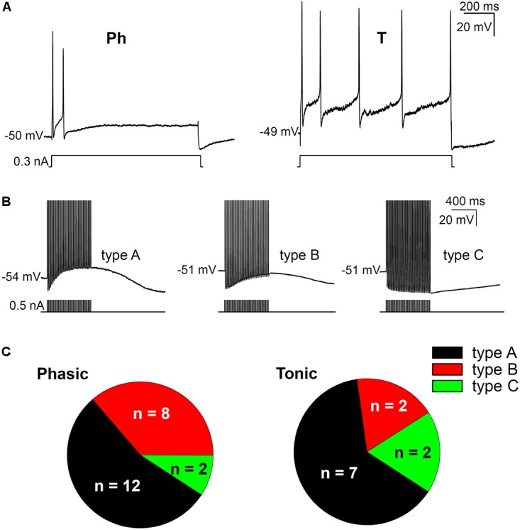
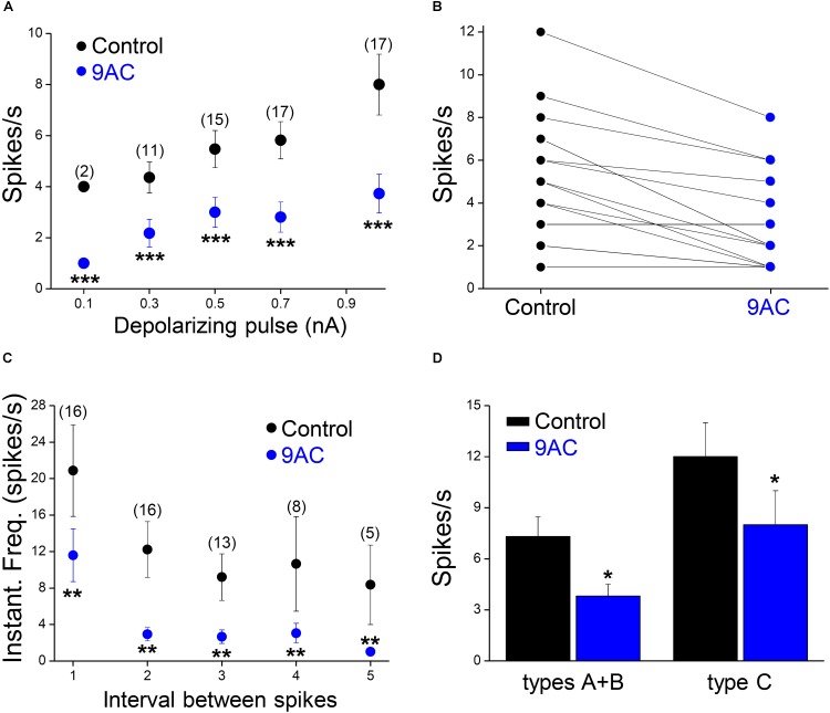
Ca-activated ion channels shape membrane excitability in response to elevations in intracellular Ca. The most extensively studied Ca-sensitive ion channels are Ca-activated K channels, whereas the physiological importance of Ca-activated Cl channels has been poorly studied. Here we show that a Ca-activated Cl currents (CaCCs) modulate repetitive firing in mouse sympathetic ganglion cells. Electrophysiological recording of mouse sympathetic neurons in an preparation of the superior cervical ganglion (SCG) identifies neurons with two different firing patterns in response to long depolarizing current pulses (1 s). Neurons classified as phasic (Ph) made up 67% of the cell population whilst the remainders were tonic (T). When a high frequency train of spikes was induced by intracellular current injection, SCG sympathetic neurons reached an afterpotential mainly dependent on the ratio of activation of two Ca-dependent currents: the K [I] and CaCC. When the I was larger, an afterhyperpolarization was the predominant afterpotential but when the CaCC was larger, an afterdepolarization (ADP) was predominant. These afterpotentials can be observed after a single action potential (AP). Ph and T neurons had similar ADPs and hence, the CaCC does not seem to determine the firing pattern (Ph or T) of these neurons. However, inhibition of Ca-activated Cl channels with anthracene-9'-carboxylic acid (9AC) selectively inhibits the ADP, reducing the firing frequency and the instantaneous frequency without affecting the characteristics of single- or first-spike firing of both Ph and T neurons. Furthermore, we found that the CaCC underlying the ADP was significantly larger in SCG neurons from males than from females. Furthermore, the CaCC ANO1/TMEM16A was more strongly expressed in male than in female SCGs. Blocking ADPs with 9AC did not modify synaptic transmission in either Ph or T neurons. We conclude that the CaCC responsible for ADPs increases repetitive firing in both Ph and T neurons, and it is more relevant in male mouse sympathetic ganglion neurons.
钙激活离子通道可根据细胞内钙离子浓度升高的情况来塑造膜兴奋性。研究最为广泛的钙敏感离子通道是钙激活钾通道,而钙激活氯通道的生理重要性却鲜少被研究。在此我们表明,一种钙激活氯电流(CaCCs)可调节小鼠交感神经节细胞的重复放电。在颈上神经节(SCG)标本中对小鼠交感神经元进行电生理记录,可识别出在响应长时间去极化电流脉冲(1秒)时具有两种不同放电模式的神经元。被归类为相位型(Ph)的神经元占细胞总数的67%,其余的为紧张型(T)。当通过细胞内电流注入诱导出高频尖峰序列时,SCG交感神经元会达到一个后电位,该后电位主要取决于两种钙依赖性电流的激活比例:钾电流[I]和CaCC。当I较大时,超极化后电位是主要的后电位,但当CaCC较大时,去极化后电位(ADP)则占主导。这些后电位可在单个动作电位(AP)后观察到。Ph和T神经元具有相似的ADP,因此,CaCC似乎并不能决定这些神经元的放电模式(Ph或T)。然而,用蒽-9'-羧酸(9AC)抑制钙激活氯通道可选择性地抑制ADP,降低放电频率和瞬时频率,而不影响Ph和T神经元的单次或首次尖峰放电特性。此外,我们发现,雄性SCG神经元中ADP所依赖的CaCC明显大于雌性。此外,钙激活氯通道ANO1/TMEM16A在雄性SCG中的表达比雌性更强。用9AC阻断ADP并不会改变Ph或T神经元中的突触传递。我们得出结论,负责ADP的CaCC会增加Ph和T神经元的重复放电,并且在雄性小鼠交感神经节神经元中更为重要。