A.I. Virtanen Institute for Molecular Sciences, University of Eastern Finland, Kuopio, Finland.
Center for Magnetic Resonance Research, Minneapolis, MN, USA.
J Cardiovasc Magn Reson. 2018 Jun 7;20(1):34. doi: 10.1186/s12968-018-0463-x.
Two days after myocardial infarction (MI), the infarct consists mostly on necrotic tissue, and the myocardium is transformed through granulation tissue to scar in two weeks after the onset of ischemia in mice. In the current work, we determined and optimized cardiovascular magnetic resonance (CMR) methods for the detection of MI size during the scar formation without contrast agents in mice.
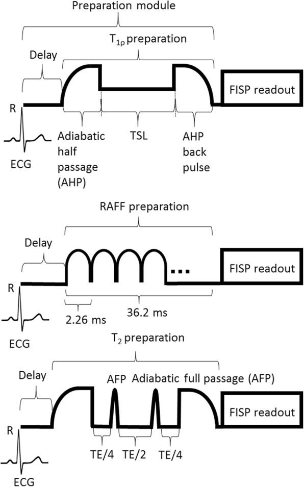
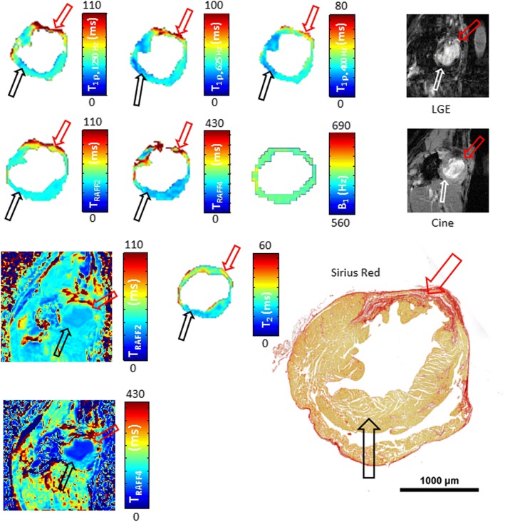
We characterized MI and remote areas with rotating frame relaxation time mapping including relaxation along fictitious field in n rotating frame (RAFFn), T and T relaxation time mappings at 1, 3, 7, and 21 days after MI. These results were compared to late gadolinium enhancement (LGE) and Sirius Red-stained histology sections, which were obtained at day 21 after MI.
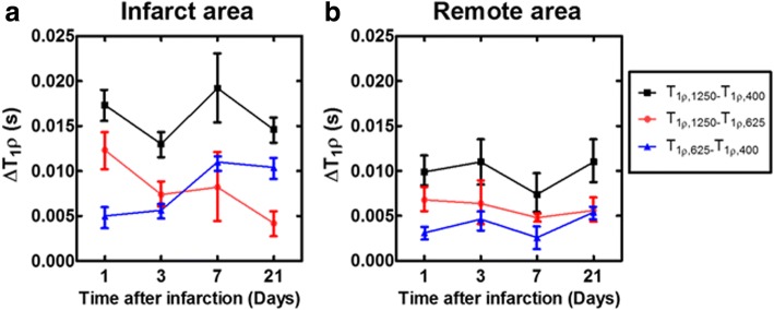
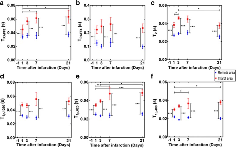
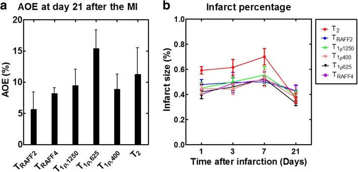
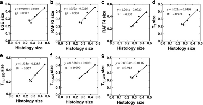
All relaxation time maps showed significant differences in relaxation time between the MI and remote area. Areas of increased signal intensities after gadolinium injection and areas with increased T relaxation time were highly correlated with the MI area determined from Sirius Red-stained histology sections (LGE: R = 0.92, P < 0.01, T: R = 0.95, P < 0.001). Infarct area determined based on T relaxation time correlated highly with Sirius Red histology sections (R = 0.97, P < 0.01). The smallest overestimation of the LGE-defined MI area was obtained for T (5.6 ± 4.2%) while for T overestimation percentage was > 9% depending on T pulse power.
T and T relaxation time maps can be used to determine accurately MI area at various time points in the mouse heart. Determination of MI size based on T relaxation time maps could be performed without contrast agents, unlike LGE, and with lower specific absorption rate compared to on-resonance T relaxation time mapping.
心肌梗死(MI)发生两天后,梗死区主要由坏死组织组成,在缺血发生后两周内,心肌通过肉芽组织转化为瘢痕。在目前的工作中,我们确定并优化了心血管磁共振(CMR)方法,用于在没有造影剂的情况下检测小鼠缺血后瘢痕形成过程中的 MI 大小。
我们通过旋转帧弛豫时间映射(包括在 n 个旋转帧中的虚构场弛豫时间映射(RAFFn))、T 和 T 弛豫时间映射来对 MI 和远程区域进行特征描述,这些成像在 MI 发生后 1、3、7 和 21 天进行。这些结果与 MI 发生后 21 天获得的钆增强延迟期(LGE)和 Sirius Red 染色组织学切片进行了比较。
所有弛豫时间图均显示 MI 和远程区域之间的弛豫时间存在显著差异。注射钆后信号强度增加的区域和 T 弛豫时间增加的区域与 Sirius Red 染色组织学切片确定的 MI 区域高度相关(LGE:R=0.92,P<0.01,T:R=0.95,P<0.001)。基于 T 弛豫时间确定的梗死面积与 Sirius Red 组织学切片高度相关(R=0.97,P<0.01)。T 确定的 LGE 定义的 MI 区域的最小高估百分比为 5.6%±4.2%,而 T 脉冲功率的高估百分比大于 9%。
T 和 T 弛豫时间图可用于在小鼠心脏的各个时间点准确确定 MI 区域。与 LGE 不同,基于 T 弛豫时间图确定 MI 大小可以在不使用造影剂的情况下进行,并且与基于共振 T 弛豫时间映射相比,SAR 更低。