Department of Internal Medicine I, University Hospital Würzburg, Würzburg, Germany.
Experimental Physics 5, University of Würzburg, Würzburg, Germany.
J Cardiovasc Magn Reson. 2022 May 9;24(1):30. doi: 10.1186/s12968-022-00864-2.
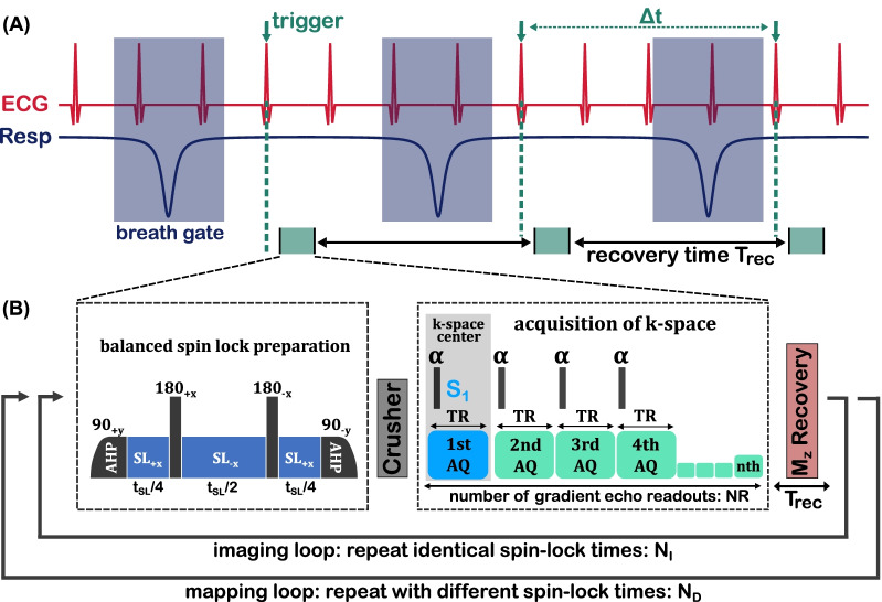
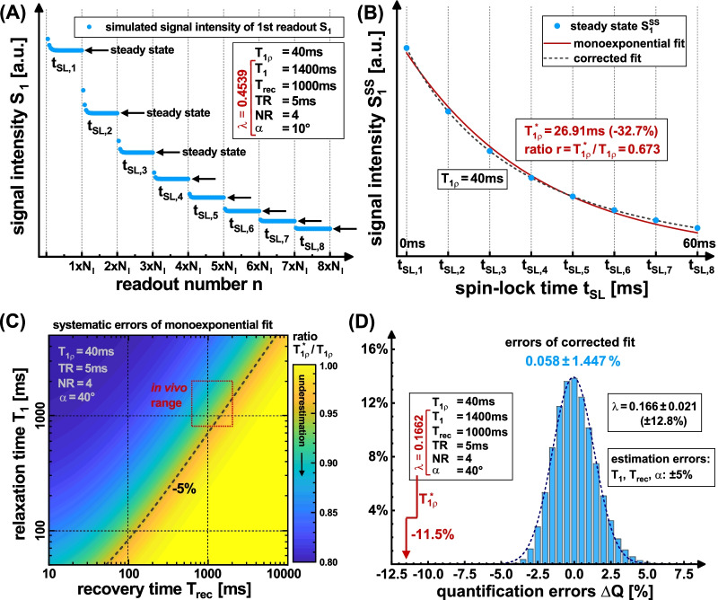
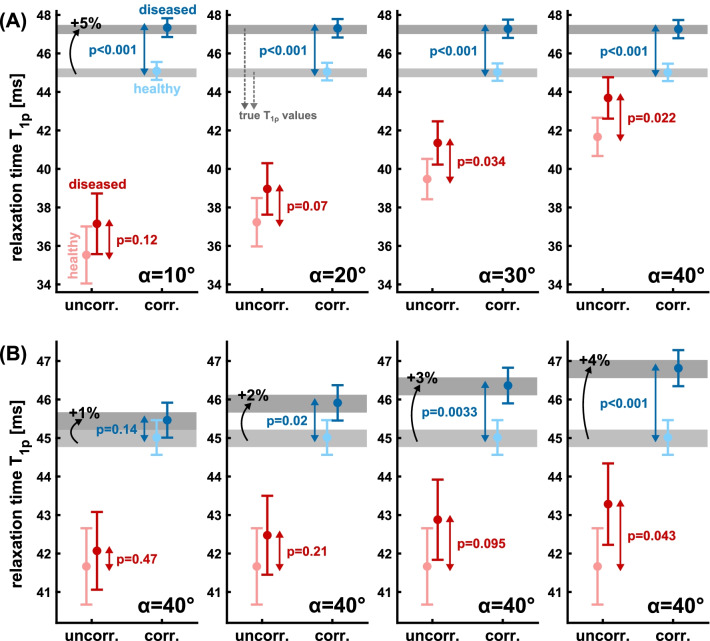
Fast and accurate T mapping in myocardium is still a major challenge, particularly in small animal models. The complex sequence design owing to electrocardiogram and respiratory gating leads to quantification errors in in vivo experiments, due to variations of the T relaxation pathway. In this study, we present an improved quantification method for T using a newly derived formalism of a T* relaxation pathway.
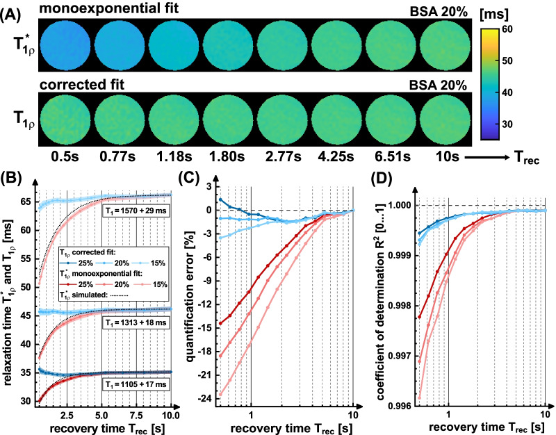
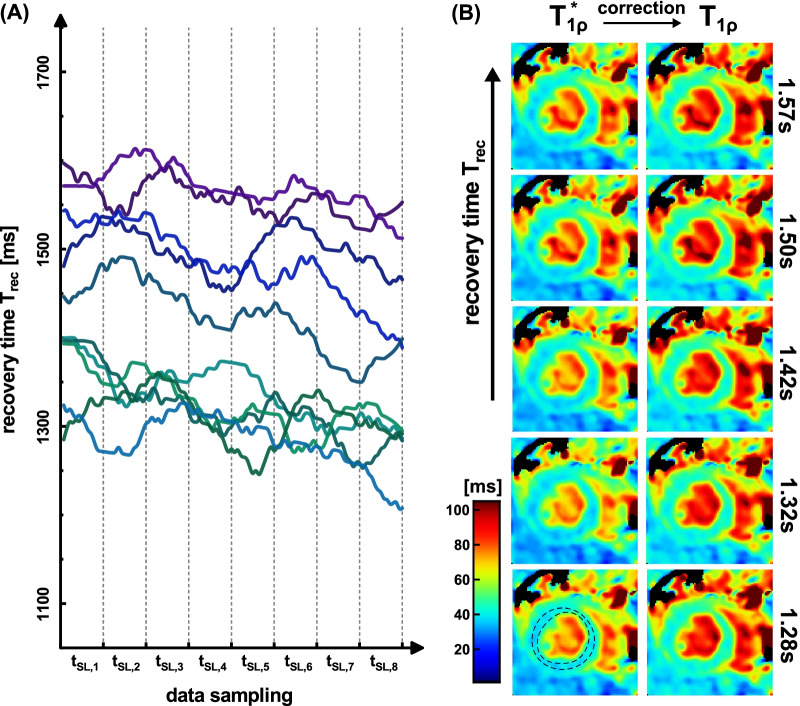
The new signal equation was derived by solving a recursion problem for spin-lock prepared fast gradient echo readouts. Based on Bloch simulations, we compared quantification errors using the common monoexponential model and our corrected model. The method was validated in phantom experiments and tested in vivo for myocardial T mapping in mice. Here, the impact of the breath dependent spin recovery time T on the quantification results was examined in detail.
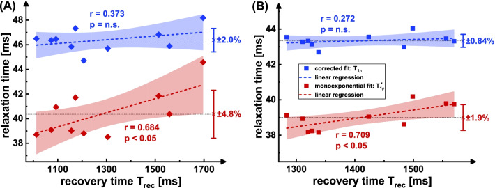
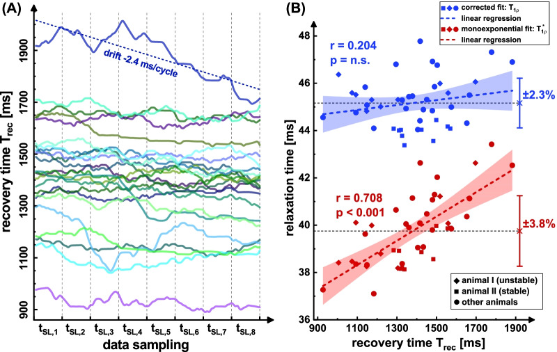
Simulations indicate that a correction is necessary, since systematically underestimated values are measured under in vivo conditions. In the phantom study, the mean quantification error could be reduced from - 7.4% to - 0.97%. In vivo, a correlation of uncorrected T with the respiratory cycle was observed. Using the newly derived correction method, this correlation was significantly reduced from r = 0.708 (p < 0.001) to r = 0.204 and the standard deviation of left ventricular T values in different animals was reduced by at least 39%.
The suggested quantification formalism enables fast and precise myocardial T quantification for small animals during free breathing and can improve the comparability of study results. Our new technique offers a reasonable tool for assessing myocardial diseases, since pathologies that cause a change in heart or breathing rates do not lead to systematic misinterpretations. Besides, the derived signal equation can be used for sequence optimization or for subsequent correction of prior study results.
快速准确的心肌 T 映射仍然是一个主要挑战,尤其是在小动物模型中。由于心电图和呼吸门控的复杂序列设计,导致 T 弛豫途径的变化,在体内实验中会导致定量误差。在这项研究中,我们提出了一种使用新导出的 T*弛豫途径形式主义来进行 T 定量的改进方法。
通过求解自旋锁定快速梯度回波读出的递归问题,推导出新的信号方程。基于布洛赫模拟,我们比较了使用常见的单指数模型和我们修正的模型的定量误差。该方法在体模实验中进行了验证,并在小鼠的心肌 T 映射中进行了体内测试。在这里,详细检查了依赖呼吸的自旋恢复时间 T 对定量结果的影响。
模拟表明,由于在体内条件下测量到系统低估的值,因此需要进行修正。在体模研究中,平均定量误差可以从-7.4%降低到-0.97%。在体内,观察到未校正 T 与呼吸周期之间存在相关性。使用新导出的校正方法,这种相关性从 r=0.708(p<0.001)显著降低到 r=0.204,并且不同动物左心室 T 值的标准差降低了至少 39%。
建议的定量形式主义使小动物在自由呼吸时能够快速准确地进行心肌 T 定量,并提高研究结果的可比性。我们的新技术为评估心肌疾病提供了一种合理的工具,因为导致心脏或呼吸率变化的病变不会导致系统的误解。此外,推导的信号方程可用于序列优化或后续校正先前的研究结果。