Department of Breast Cancer Center, Shandong Cancer Hospital Affiliated to Shandong University, Jinan, China.
Shandong Academy of Medical Sciences, Jinan, China.
Cancer Res Treat. 2019 Apr;51(2):483-492. doi: 10.4143/crt.2018.062. Epub 2018 Jun 11.
The purpose of this study was to detect the lymphatic drainage pattern of internal mammary area and verify the concept of internal mammary sentinel lymph node (IM-SLN) in breast.
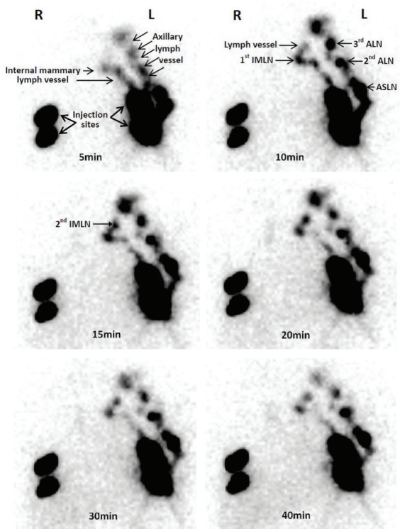
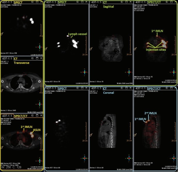
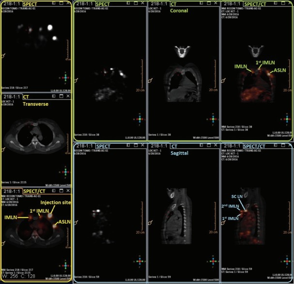
A small particle radiotracer (99mTc-Dextran 40) was prepared and tested. 99mTc-Dextran 40 was injected into intraparenchyma at the sound breast by a modified radiotracer injection technique. Subsequently, dynamic single-photon emission computed tomography (SPECT), computed tomography (CT), and SPECT/CT combination images were performed to identify the radioactive lymph vessels and internal mammary lymph nodes (IMLNs). The direction of lymph drainage and the location of the IMLNs were identified in the SPECT/CT imaging.
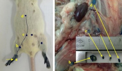
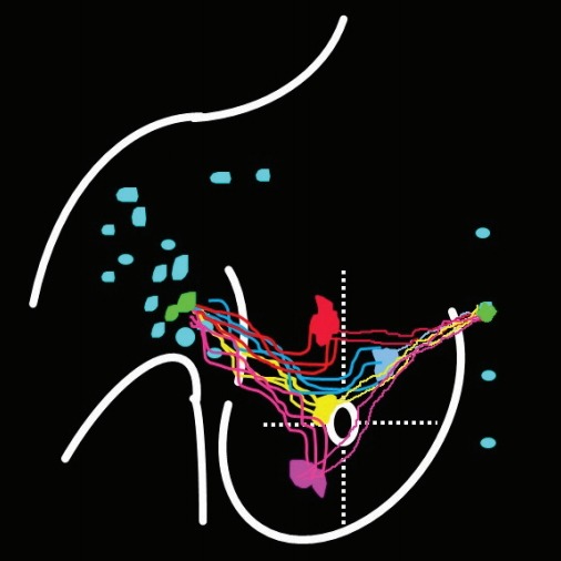
The radiochemical purity of 99mTc-Dextran 40 was > 95%. 99mTc-Dextran 40 could drainage into first, second, and third lymph node and the radioactive lymph node could be detected by the γ detector in the animal experiment. After 99mTc-Dextran 40 injecting into intraparenchyma, 50.0% cases (15/30) were identified the drainage lymphatic vessels and radioactive IMLNs by SPECT. The drainage lymphatic vessel was found from injection point to the first IMLN (IM-SLN) after 10.5±0.35 minutes radiotracer injection, and then 99mTc-Dextran 40 was accumulated into the IM-SLN. The combination imaging of SPECT/CT showed the second IMLN received the lymph drainage from the IM-SLN. The lymphatic drainage was step by step in the internal mammary area.
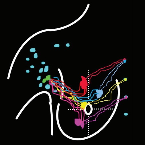
The lymph was identified to drain from different regions of the breast to IM-SLN, and then outward from IM-SLN to other IMLN consecutively. It demonstrated the concept of the IM-SLN and provided more evidences for the application of internal mammary sentinel lymph node biopsy.
本研究旨在探测内乳区的淋巴引流模式,并验证乳腺内乳前哨淋巴结(IM-SLN)的概念。
制备并测试了一种小颗粒放射性示踪剂(99mTc-Dextran 40)。通过改良的放射性示踪剂注射技术,将 99mTc-Dextran 40 注入乳腺实质内。随后,进行动态单光子发射计算机断层扫描(SPECT)、计算机断层扫描(CT)和 SPECT/CT 组合图像,以识别放射性淋巴管和内乳淋巴结(IMLN)。在 SPECT/CT 成像中确定了淋巴引流的方向和 IMLN 的位置。
99mTc-Dextran 40 的放射化学纯度>95%。99mTc-Dextran 40 可引流至第一、第二和第三淋巴结,并且可在动物实验中通过γ探测器检测到放射性淋巴结。在 99mTc-Dextran 40 注入乳腺实质后,50.0%(15/30)的病例通过 SPECT 识别出引流淋巴管和放射性 IMLN。在放射性示踪剂注射后 10.5±0.35 分钟,发现引流淋巴管从注射点到第一内乳前哨淋巴结(IM-SLN),然后 99mTc-Dextran 40 积聚到 IM-SLN 中。SPECT/CT 组合图像显示第二内乳淋巴结接收来自 IM-SLN 的淋巴引流。内乳区域的淋巴引流是逐步进行的。
淋巴被识别从乳房的不同区域引流到 IM-SLN,然后从 IM-SLN 向外连续引流到其他 IMLN。这证明了 IM-SLN 的概念,并为内乳前哨淋巴结活检的应用提供了更多证据。