The Department of Plastic and Reconstructive Surgery, Tel Aviv Sourasky Medical Center, Sackler Faculty of Medicine, Tel Aviv University, Tel Aviv, Israel.
The Cancer Immunotherapy lab, The Neurosurgery department, Tel Aviv Sourasky Medical Center, Sackler Faculty of Medicine, Tel Aviv University, Tel Aviv, Israel.
Cell Death Dis. 2018 Jun 11;9(6):695. doi: 10.1038/s41419-018-0702-y.
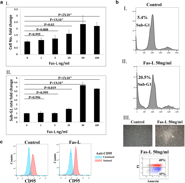
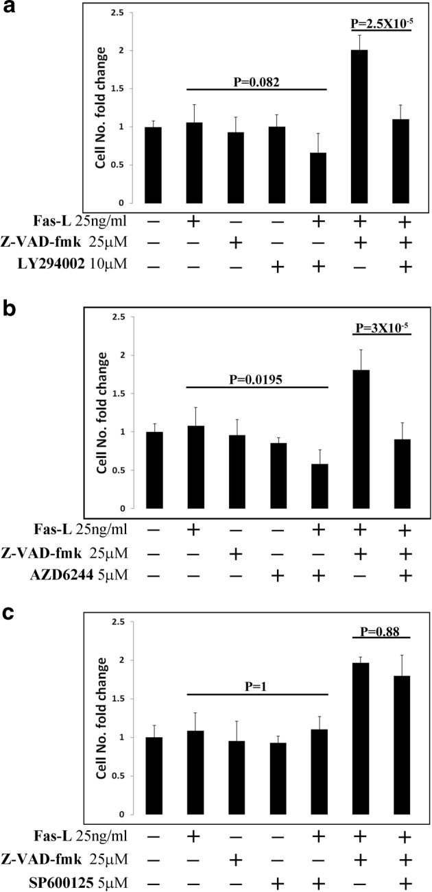
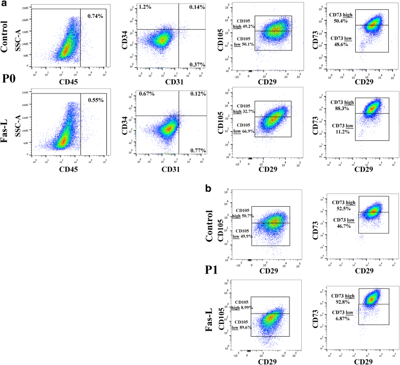
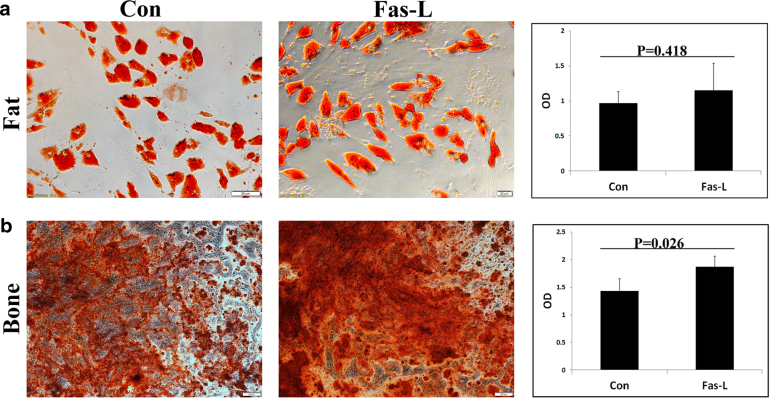
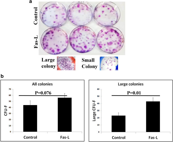
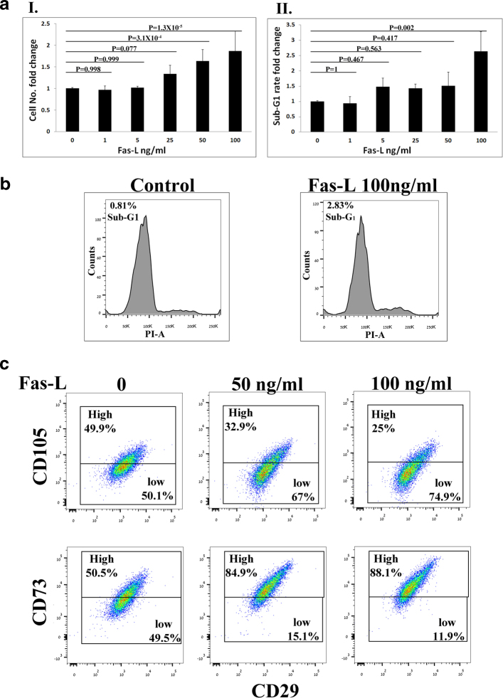
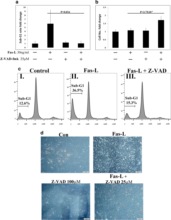
Fas-L is a TNF family member known to trigger cell death. It has recently become evident that Fas-L can transduce also non-apoptotic signals. Mesenchymal stem cells (MSCs) are multipotent cells that are derived from various adult tissues. Although MSCs from different tissues display common properties they also display tissue-specific characteristics. Previous works have demonstrated massive apoptosis following Fas-L treatment of bone marrow-derived MSCs both in vitro and following their administration in vivo. We therefore set to examine Fas-L-induced responses in adipose-derived stem cells (ASCs). Human ASCs were isolated from lipoaspirates and their reactivity to Fas-L treatment was examined. ASCs responded to Fas-L by simultaneous apoptosis and proliferation, which yielded a net doubling of cell quantities and a phenotypic shift, including reduced expression of CD105 and increased expression of CD73, in association with increased bone differentiation potential. Treatment of freshly isolated ASCs led to an increase in large colony forming unit fibroblasts, likely produced by early stem cell progenitor cells. Fas-L-induced apoptosis and proliferation signaling were found to be independent as caspase inhibition attenuated Fas-L-induced apoptosis without impacting proliferation, whereas inhibition of PI3K and MEK, but not of JNK, attenuated Fas-L-dependent proliferation, but not apoptosis. Thus, Fas-L signaling in ASCs leads to their expansion and phenotypic shift toward a more potent stem cell state. We speculate that these reactions ensure the survival of ASC progenitor cells encountering Fas-L-enriched environments during tissue damage and inflammation and may also enhance ASC survival following their administration in vivo.
Fas-L 是 TNF 家族的一员,已知能触发细胞死亡。最近的研究表明,Fas-L 还可以传递非凋亡信号。间充质干细胞(MSCs)是多能细胞,源自各种成人组织。虽然来自不同组织的 MSCs 具有共同的特性,但它们也具有组织特异性特征。以前的研究表明,Fas-L 处理骨髓来源的 MSCs 会导致大量细胞凋亡,无论是在体外还是在体内给药后。因此,我们着手研究 Fas-L 在脂肪来源的干细胞(ASCs)中诱导的反应。从脂肪抽吸物中分离出人 ASC,并检查 Fas-L 处理对其的反应。ASCs 通过同时发生凋亡和增殖来响应 Fas-L,这导致细胞数量净加倍和表型转变,包括 CD105 表达减少和 CD73 表达增加,同时骨分化潜能增加。对新鲜分离的 ASC 的处理导致大集落形成单位成纤维细胞数量增加,这可能是由早期干细胞祖细胞产生的。发现 Fas-L 诱导的凋亡和增殖信号是独立的,因为 caspase 抑制剂减弱 Fas-L 诱导的凋亡而不影响增殖,而 PI3K 和 MEK 的抑制,但不是 JNK 的抑制,减弱 Fas-L 依赖性增殖,但不减弱凋亡。因此,Fas-L 信号在 ASCs 中导致其扩增和表型向更具潜能的干细胞状态转变。我们推测,这些反应确保了在组织损伤和炎症过程中遇到 Fas-L 富集环境的 ASC 祖细胞的存活,并可能在体内给药后也增强 ASC 的存活。