• 文献检索
  • 文档翻译
  • 深度研究
  • 学术资讯
  • Suppr Zotero 插件Zotero 插件
  • 邀请有礼
  • 套餐&价格
  • 历史记录
应用&插件
Suppr Zotero 插件Zotero 插件浏览器插件Mac 客户端Windows 客户端微信小程序
定价
高级版会员购买积分包购买API积分包
服务
文献检索文档翻译深度研究API 文档MCP 服务
关于我们
关于 Suppr公司介绍联系我们用户协议隐私条款
关注我们

Suppr 超能文献

核心技术专利:CN118964589B侵权必究
粤ICP备2023148730 号-1Suppr @ 2026

文献检索

告别复杂PubMed语法,用中文像聊天一样搜索,搜遍4000万医学文献。AI智能推荐,让科研检索更轻松。

立即免费搜索

文件翻译

保留排版,准确专业,支持PDF/Word/PPT等文件格式,支持 12+语言互译。

免费翻译文档

深度研究

AI帮你快速写综述,25分钟生成高质量综述,智能提取关键信息,辅助科研写作。

立即免费体验

颈动脉易损斑块与急性缺血性脑卒中患者循环白细胞相关:一项基于超声造影的临床研究。

Carotid vulnerable plaques are associated with circulating leukocytes in acute ischemic stroke patients: an clinical study based on contrast-enhanced ultrasound.

机构信息

Department of Ultrasound, Shanghai General Hospital, Shanghai Jiaotong University School of Medicine, Shanghai, China.

Department of Echocardiography, Shanghai General Hospital, Shanghai Jiaotong University School of Medicine, Shanghai, China.

出版信息

Sci Rep. 2018 Jun 11;8(1):8849. doi: 10.1038/s41598-018-27260-0.

DOI:10.1038/s41598-018-27260-0
PMID:29892086
原文链接:https://pmc.ncbi.nlm.nih.gov/articles/PMC5995867/
Abstract

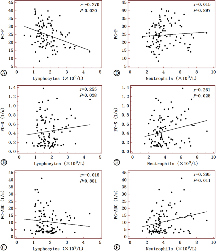
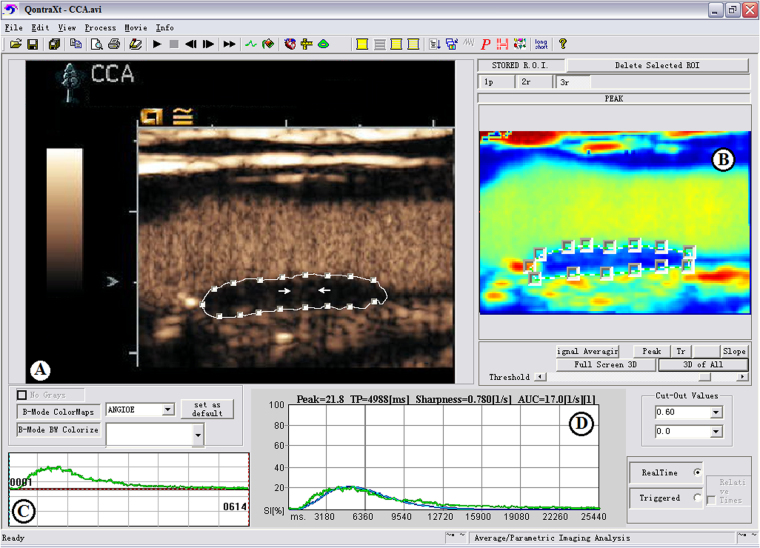
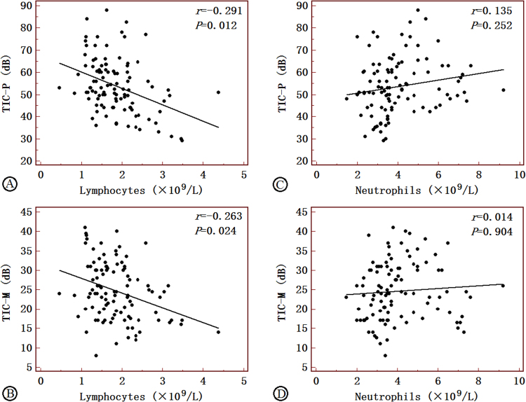
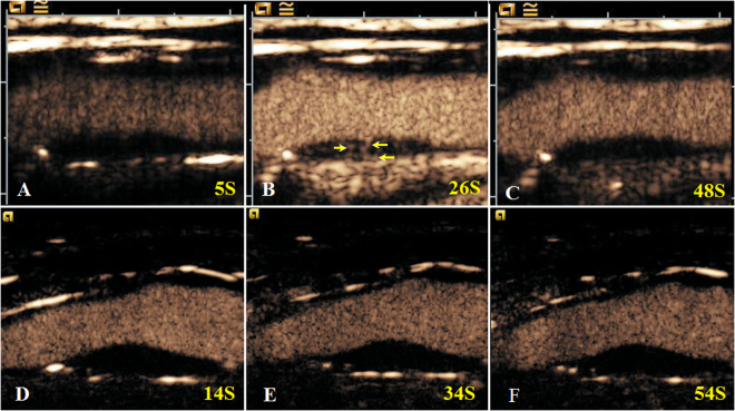
Inflammatory activity plays a central role in the development of carotid rupture-vulnerable atherosclerotic plaques, which is one of the major contributors to acute ischemic stroke. Our objective was to characterize carotid intraplaque neovascularizations (INP) using contrast-enhanced ultrasound (CEUS) and evaluate plaque burden through exploring the relationship between INP and cell count of peripheral leukocytes. Sixty-two patients with large artery atherosclerosis (LAA) were enrolled in this study. CEUS was performed to characterize the carotid artery plaques. The correlations between the CEUS imaging features of carotid plaques and leukocyte counts were investigated. The results showed that the characteristic parameters derived from CEUS, including peak of time-intensity curve (TIC-P), mean of time-intensity curve (TIC-M), peak (FC-P), sharpness (FC-S) and area under the curve (FC-AUC) compared with the control group, were all increased in the stroke group. TIC-P, TIC-M and FC-P were negatively related to lymphocytes, respectively. FC-S and FC-AUC were positively correlated with neutrophils, respectively. Our study indicated carotid INP was closely related to the peripheral leukocytes count. CEUS may serve as a useful tool to predict vulnerability of plaque.

摘要

炎症活动在颈动脉易破裂斑块的形成中起着核心作用,而颈动脉易破裂斑块是急性缺血性脑卒中的主要原因之一。本研究旨在通过探讨颈动脉斑块内新生血管(INP)与外周血白细胞计数之间的关系,利用对比增强超声(CEUS)来描述颈动脉斑块内的新生血管。本研究纳入了 62 例大动脉粥样硬化(LAA)患者,进行 CEUS 以描述颈动脉斑块的特征。研究了颈动脉斑块的 CEUS 成像特征与白细胞计数之间的相关性。结果表明,与对照组相比,CEUS 特征参数,包括时间强度曲线(TIC)峰值(TIC-P)、TIC 均值(TIC-M)、峰值(FC-P)、锐利度(FC-S)和曲线下面积(FC-AUC)在卒中组中均增加。TIC-P、TIC-M 和 FC-P 分别与淋巴细胞呈负相关。FC-S 和 FC-AUC 分别与中性粒细胞呈正相关。我们的研究表明颈动脉 INP 与外周血白细胞计数密切相关。CEUS 可能是一种预测斑块易损性的有用工具。

https://cdn.ncbi.nlm.nih.gov/pmc/blobs/c202/5995867/ddb475168c23/41598_2018_27260_Fig5_HTML.jpg
https://cdn.ncbi.nlm.nih.gov/pmc/blobs/c202/5995867/d02f7d015dc3/41598_2018_27260_Fig1_HTML.jpg
https://cdn.ncbi.nlm.nih.gov/pmc/blobs/c202/5995867/eb118cca7fbf/41598_2018_27260_Fig2_HTML.jpg
https://cdn.ncbi.nlm.nih.gov/pmc/blobs/c202/5995867/8bff682e9a18/41598_2018_27260_Fig3_HTML.jpg
https://cdn.ncbi.nlm.nih.gov/pmc/blobs/c202/5995867/ca9fa404785b/41598_2018_27260_Fig4_HTML.jpg
https://cdn.ncbi.nlm.nih.gov/pmc/blobs/c202/5995867/ddb475168c23/41598_2018_27260_Fig5_HTML.jpg

相似文献

1
Carotid vulnerable plaques are associated with circulating leukocytes in acute ischemic stroke patients: an clinical study based on contrast-enhanced ultrasound.颈动脉易损斑块与急性缺血性脑卒中患者循环白细胞相关:一项基于超声造影的临床研究。
Sci Rep. 2018 Jun 11;8(1):8849. doi: 10.1038/s41598-018-27260-0.
2
Relation between carotid vulnerable plaques and peripheral leukocyte: a case-control study of comparison utilizing multi-parametric contrast-enhanced ultrasound.颈动脉易损斑块与外周血白细胞的关系:多参数对比增强超声的病例对照研究。
BMC Med Imaging. 2019 Aug 23;19(1):74. doi: 10.1186/s12880-019-0374-9.
3
Evaluation of Carotid Plaque Rupture and Neovascularization by Contrast-Enhanced Ultrasound Imaging: an Exploratory Study Based on Histopathology.基于组织病理学的颈动脉斑块破裂与新生血管的超声造影评估:一项探索性研究。
Transl Stroke Res. 2021 Feb;12(1):49-56. doi: 10.1007/s12975-020-00825-w. Epub 2020 Jun 29.
4
Contrast-Enhanced Ultrasound to Assess Carotid Intraplaque Neovascularization.对比增强超声评估颈动脉斑块内新生血管。
Ultrasound Med Biol. 2020 Mar;46(3):466-478. doi: 10.1016/j.ultrasmedbio.2019.10.020. Epub 2019 Nov 29.
5
Utility of Complementary Magnetic Resonance Plaque Imaging and Contrast-Enhanced Ultrasound to Detect Carotid Vulnerable Plaques.补充磁共振斑块成像和对比增强超声检测颈动脉易损斑块的效用。
J Am Heart Assoc. 2019 Apr 16;8(8):e011302. doi: 10.1161/JAHA.118.011302.
6
Carotid Plaque Neovascularization Detected With Superb Microvascular Imaging Ultrasound Without Using Contrast Media.颈动脉斑块内新生血管的超声造影检测技术。
Stroke. 2019 Nov;50(11):3121-3127. doi: 10.1161/STROKEAHA.119.025496. Epub 2019 Sep 12.
7
Plaque Elasticity and Intraplaque Neovascularisation on Carotid Artery Ultrasound: A Comparative Histological Study.颈动脉超声斑块弹性和斑块内新生血管化:一项对比组织学研究。
Eur J Vasc Endovasc Surg. 2021 Sep;62(3):358-366. doi: 10.1016/j.ejvs.2021.05.026.
8
Contrast-enhanced ultrasound evaluation of plaque vulnerability and the relationship between peripheral blood leukocytes.超声造影评价斑块易损性与外周血白细胞的关系
Clin Hemorheol Microcirc. 2024;87(2):187-197. doi: 10.3233/CH-232034.
9
[Value of Superb Micro-vascular Imaging in Diagnosing Carotid Artery Vulnerable Plaque].[超微血管成像在诊断颈动脉易损斑块中的价值]
Zhongguo Yi Xue Ke Xue Yuan Xue Bao. 2018 Aug 30;40(4):444-449. doi: 10.3881/j.issn.1000-503X.10424.
10
Contrast-enhanced ultrasound perfusion patterns and serum lipid signatures of vulnerable carotid artery plaque in predicting stroke: A cohort study of carotid stenosis in Chinese patients.超声造影灌注模式及血清脂质特征对易损性颈动脉斑块预测卒中的价值:一项中国患者颈动脉狭窄队列研究
Clin Hemorheol Microcirc. 2020;75(3):349-359. doi: 10.3233/CH-190804.

引用本文的文献

1
Predictive value of contrast-enhanced carotid ultrasound features for stroke risk: a systematic review and meta-analysis.对比增强型颈动脉超声特征对卒中风险的预测价值:一项系统评价和荟萃分析
Front Neurol. 2025 May 16;16:1487850. doi: 10.3389/fneur.2025.1487850. eCollection 2025.
2
Assessment of Carotid Plaque Enhancement on Contrast-Enhanced Ultrasound as a Predictor for Severe Coronary Artery Disease.对比增强超声评估颈动脉斑块强化作为严重冠状动脉疾病的预测指标
Indian J Radiol Imaging. 2024 Feb 23;34(3):460-468. doi: 10.1055/s-0044-1779522. eCollection 2024 Jul.
3
Research Advance of Chinese Medicine in Treating Atherosclerosis: Focus on Lipoprotein-Associated Phospholipase A2.

本文引用的文献

1
Impact of Fiber Structure on the Material Stability and Rupture Mechanisms of Coronary Atherosclerotic Plaques.纤维结构对冠状动脉粥样硬化斑块材料稳定性及破裂机制的影响
Ann Biomed Eng. 2017 Jun;45(6):1462-1474. doi: 10.1007/s10439-017-1827-3. Epub 2017 Mar 30.
2
Circulating CD14+ and CD14CD16- classical monocytes are reduced in patients with signs of plaque neovascularization in the carotid artery.在有颈动脉斑块新生血管形成迹象的患者中,循环中的CD14+和CD14CD16-经典单核细胞数量减少。
Atherosclerosis. 2016 Dec;255:171-178. doi: 10.1016/j.atherosclerosis.2016.10.004. Epub 2016 Oct 6.
3
The therapeutic effect of bevacizumab on plaque neovascularization in a rabbit model of atherosclerosis during contrast-enhanced ultrasonography.
中药治疗动脉粥样硬化的研究进展:关注脂蛋白相关磷脂酶 A2。
Chin J Integr Med. 2024 Mar;30(3):277-288. doi: 10.1007/s11655-023-3611-6. Epub 2023 Dec 7.
4
Advances in Noninvasive Carotid Wall Imaging with Ultrasound: A Narrative Review.超声无创颈动脉壁成像的进展:一篇叙述性综述
J Clin Med. 2022 Oct 20;11(20):6196. doi: 10.3390/jcm11206196.
5
The Construction and Analysis of Immune Infiltration and Competing Endogenous RNA Network in Acute Ischemic Stroke.急性缺血性卒中免疫浸润与竞争性内源性RNA网络的构建及分析
Front Aging Neurosci. 2022 May 17;14:806200. doi: 10.3389/fnagi.2022.806200. eCollection 2022.
6
Per-pass analysis of acute ischemic stroke clots: impact of stroke etiology on extracted clot area and histological composition.单次通过分析急性缺血性脑卒中血栓:发病机制对提取血栓面积和组织学成分的影响。
J Neurointerv Surg. 2021 Dec;13(12):1111-1116. doi: 10.1136/neurintsurg-2020-016966. Epub 2020 Dec 9.
7
CT imaging features of carotid artery plaque vulnerability.颈动脉斑块易损性的CT成像特征
Ann Transl Med. 2020 Oct;8(19):1261. doi: 10.21037/atm-2020-cass-13.
8
Contrast imaging ultrasound for the detection and characterization of carotid vulnerable plaque.超声造影成像用于检测和鉴定颈动脉易损斑块
Cardiovasc Diagn Ther. 2020 Aug;10(4):965-981. doi: 10.21037/cdt.2020.01.08.
9
Assessment of Carotid Intraplaque Neovascularization Using Superb Microvascular Imaging in High Risk of Stroke Individuals: Results From a Community-Based Study.使用超高分辨率微血管成像技术评估脑卒中高危个体的颈动脉斑块内新生血管形成:一项基于社区研究的结果
Front Neurol. 2019 Nov 7;10:1146. doi: 10.3389/fneur.2019.01146. eCollection 2019.
10
Neprilysin Inhibitor-Angiotensin II Receptor Blocker Combination Therapy (Sacubitril/valsartan) Suppresses Atherosclerotic Plaque Formation and Inhibits Inflammation in Apolipoprotein E- Deficient Mice.奈必洛尔-血管紧张素受体阻滞剂联合治疗(沙库巴曲缬沙坦)抑制载脂蛋白 E 缺陷小鼠动脉粥样硬化斑块形成并抑制炎症。
Sci Rep. 2019 Apr 24;9(1):6509. doi: 10.1038/s41598-019-42994-1.
贝伐单抗对兔动脉粥样硬化模型斑块新生血管在超声造影检查中的治疗效果。
Sci Rep. 2016 Jul 25;6:30417. doi: 10.1038/srep30417.
4
Evaluating arterial stiffness in type 2 diabetes patients using ultrasonic radiofrequency.使用超声射频评估2型糖尿病患者的动脉僵硬度。
J Huazhong Univ Sci Technolog Med Sci. 2016 Jun;36(3):442-448. doi: 10.1007/s11596-016-1606-7. Epub 2016 Jul 5.
5
Assessment of the arterial stiffness in patients with acute ischemic stroke using longitudinal elasticity modulus measurements obtained with Shear Wave Elastography.使用剪切波弹性成像获得的纵向弹性模量测量值评估急性缺血性中风患者的动脉僵硬度。
Med Ultrason. 2016 Jun;18(2):182-9. doi: 10.11152/mu.2013.2066.182.wav.
6
Reduction of Circulating HLA-DR T Cell Levels Correlates With Increased Carotid Intraplaque Neovascularization and Atherosclerotic Burden.循环中HLA-DR T细胞水平的降低与颈动脉斑块内新生血管形成增加及动脉粥样硬化负担相关。
JACC Cardiovasc Imaging. 2016 Oct;9(10):1231-1233. doi: 10.1016/j.jcmg.2015.10.010. Epub 2016 Jan 6.
7
Imaging inflammation and neovascularization in atherosclerosis: clinical and translational molecular and structural imaging targets.动脉粥样硬化中炎症与新生血管形成的影像学:临床及转化医学中的分子与结构影像学靶点
Curr Opin Cardiol. 2015 Nov;30(6):671-80. doi: 10.1097/HCO.0000000000000226.
8
Markers of inflammation associated with plaque progression and instability in patients with carotid atherosclerosis.与颈动脉粥样硬化患者斑块进展和不稳定性相关的炎症标志物。
Mediators Inflamm. 2015;2015:718329. doi: 10.1155/2015/718329. Epub 2015 Apr 16.
9
Anti-inflammatory and antiatherogenic effects of the NLRP3 inflammasome inhibitor arglabin in ApoE2.Ki mice fed a high-fat diet.高脂饮食喂养的载脂蛋白 E2. 敲除小鼠中 NLRP3 炎性小体抑制剂 arglabin 的抗炎和抗动脉粥样硬化作用。
Circulation. 2015 Mar 24;131(12):1061-70. doi: 10.1161/CIRCULATIONAHA.114.013730. Epub 2015 Jan 22.
10
Evaluation of ventricular-vascular coupling in patients with type 2 diabetes mellitus using 2-dimensional speckle tracking imaging.使用二维斑点追踪成像评估2型糖尿病患者的心室-血管耦合
J Huazhong Univ Sci Technolog Med Sci. 2014 Dec;34(6):929-934. doi: 10.1007/s11596-014-1376-z. Epub 2014 Dec 6.